

Mex3c regulates insulin-like growth factor 1 (IGF1) expression and promotes postnatal growth
- PMID: 22357625
- PMCID: PMC3327323
- DOI: 10.1091/mbc.E11-11-0960
Mex3c regulates insulin-like growth factor 1 (IGF1) expression and promotes postnatal growth
Abstract
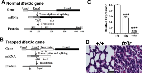
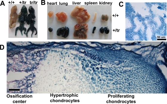
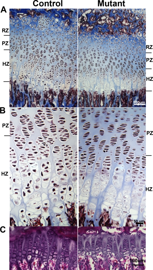
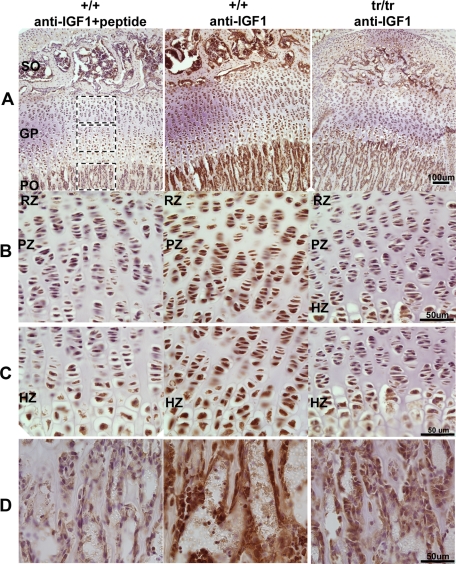
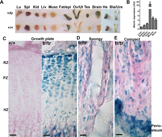
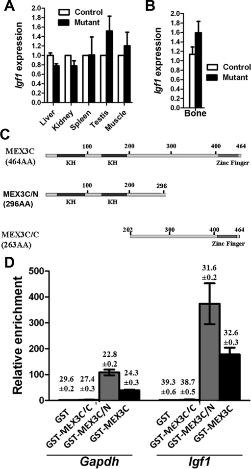
Insulin-like growth factor 1 (IGF1) mediates the growth-promoting activities of growth hormone. How Igf1 expression is regulated posttranscriptionally is unclear. Caenorhabditis elegans muscle excess 3 (MEX-3) is involved in cell fate specification during early embryonic development through regulating mRNAs involved in specifying cell fate. The function of its mammalian homologue, MEX3C, is unknown. Here we show that MEX3C deficiency in Mex3c homozygous mutant mice causes postnatal growth retardation and background-dependent perinatal lethality. Hypertrophy of chondrocytes in growth plates is significantly impaired. Circulating and bone local production of IGF1 are both decreased in mutant mice. Mex3c mRNA is strongly expressed in the testis and the brain, and highly expressed in resting and proliferating chondrocytes of the growth plates. MEX3C is able to enrich multiple mRNA species from tissue lysates, including Igf1. Igf1 expression in bone is decreased at the protein level but not at the mRNA level, indicating translational/posttranslational regulation. We propose that MEX3C protein plays an important role in enhancing the translation of Igf1 mRNA, which explains the perinatal lethality and growth retardation observed in MEX3C-deficient mice.
Figures
Similar articles
-
Mex3c mutation reduces adiposity and increases energy expenditure.Mol Cell Biol. 2012 Nov;32(21):4350-62. doi: 10.1128/MCB.00452-12. Epub 2012 Aug 27. Mol Cell Biol. 2012. PMID: 22927639 Free PMC article.
-
IGF1 regulation of BOULE and CDC25A transcripts via a testosterone-independent pathway in spermatogenesis of adult mice.Reprod Biol. 2015 Mar;15(1):48-55. doi: 10.1016/j.repbio.2014.10.003. Epub 2014 Oct 22. Reprod Biol. 2015. PMID: 25726377
-
Evidence supporting dual, IGF-I-independent and IGF-I-dependent, roles for GH in promoting longitudinal bone growth.J Endocrinol. 2004 Feb;180(2):247-55. doi: 10.1677/joe.0.1800247. J Endocrinol. 2004. PMID: 14765976
-
Oocyte-Specific Expression of Mouse MEX3C652AA in the Ovary and Its Potential Role in Regulating Maternal Fos mRNA.Biol Reprod. 2016 May;94(5):115. doi: 10.1095/biolreprod.115.136630. Epub 2016 Apr 6. Biol Reprod. 2016. PMID: 27053362
-
Effects of Eleutherococcus Extract Mixture on Endochondral Bone Formation in Rats.Int J Mol Sci. 2019 Mar 12;20(5):1253. doi: 10.3390/ijms20051253. Int J Mol Sci. 2019. PMID: 30871109 Free PMC article.
Cited by
-
Oncogenic Potential of the Dual-Function Protein MEX3A.Biology (Basel). 2021 May 7;10(5):415. doi: 10.3390/biology10050415. Biology (Basel). 2021. PMID: 34067172 Free PMC article. Review.
-
Post-transcriptional control of a stemness signature by RNA-binding protein MEX3A regulates murine adult neurogenesis.Nat Commun. 2023 Jan 23;14(1):373. doi: 10.1038/s41467-023-36054-6. Nat Commun. 2023. PMID: 36690670 Free PMC article.
-
Identification of hMex-3A and its effect on human bladder cancer cell proliferation.Oncotarget. 2017 May 22;8(37):61215-61225. doi: 10.18632/oncotarget.18050. eCollection 2017 Sep 22. Oncotarget. 2017. PMID: 28977858 Free PMC article.
-
Construction of a Mex3c Gene-Deficient Mouse Model to Study C-FOS Expression in Hypothalamic Nuclei and Observe Morphological Differences in Embryonic Neural Tube Development.Med Sci Monit. 2020 Nov 16;26:e927334. doi: 10.12659/MSM.927334. Med Sci Monit. 2020. PMID: 33191393 Free PMC article.
-
Examination of bioenergetic function in the inner mitochondrial membrane peptidase 2-like (Immp2l) mutant mice.Redox Biol. 2014;2:1008-15. doi: 10.1016/j.redox.2014.08.006. Epub 2014 Aug 28. Redox Biol. 2014. PMID: 25460737 Free PMC article.
References
-
- Agoulnik AI, Lu B, Zhu Q, Truong C, Ty MT, Arango N, Chada KK, Bishop CE. A novel gene, Pog, is necessary for primordial germ cell proliferation in the mouse and underlies the germ cell deficient mutation, gcd. Hum Mol Genet. 2002;11:3047–3053. - PubMed
-
- Beddington RS, Morgernstern J, Land H, Hogan A. An in situ transgenic enzyme marker for the midgestation mouse embryo and the visualization of inner cell mass clones during early organogenesis. Development. 1989;106:37–46. - PubMed
-
- Buchet-Poyau K, Courchet J, Hir HL, Seraphin B, Scoazec J-Y, Duret L, Domon-Dell C, Freund J-N, Billaud M. Identification and characterization of human Mex-3 proteins, a novel family of evolutionarily conserved RNA-binding proteins differentially localized to processing bodies. Nucl Acids Res. 2007;35:1289–1300. - PMC - PubMed
-
- Ciosk R, DePalma M, Priess JR. Translational regulators maintain totipotency in the Caenorhabditis elegans germline. Science. 2006;311:851–853. - PubMed
Publication types
MeSH terms
Substances
Associated data
- Actions
Grants and funding
LinkOut - more resources
Full Text Sources
Molecular Biology Databases
Miscellaneous
