. 2012 Apr 1;188(7):3351-63.
doi: 10.4049/jimmunol.1102863. Epub 2012 Feb 22.

Francisella tularensis inhibits the intrinsic and extrinsic pathways to delay constitutive apoptosis and prolong human neutrophil lifespan

Affiliations

Francisella tularensis inhibits the intrinsic and extrinsic pathways to delay constitutive apoptosis and prolong human neutrophil lifespan

Justin T Schwartz et al. J Immunol. .

Abstract

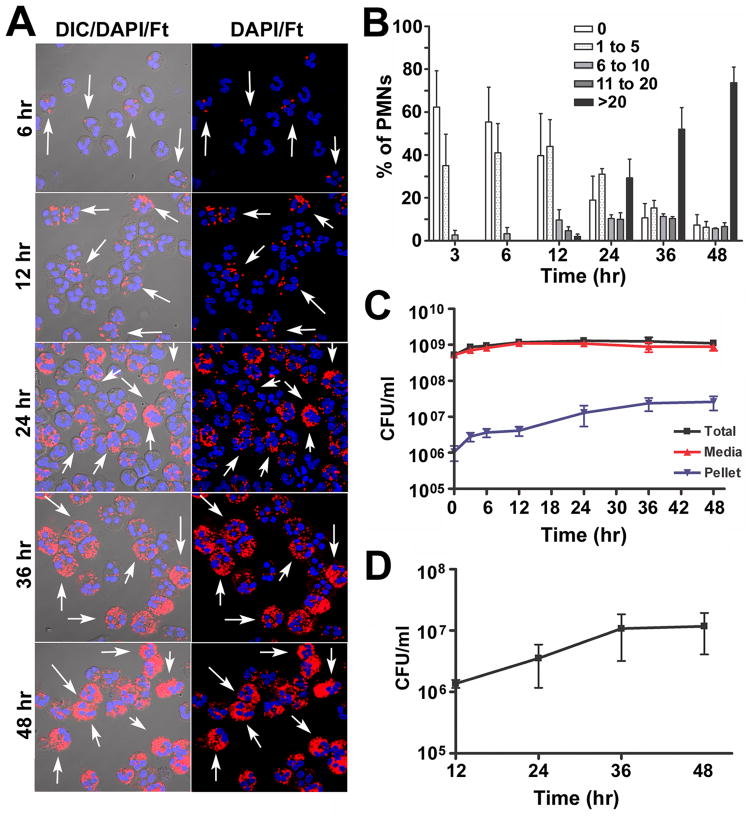
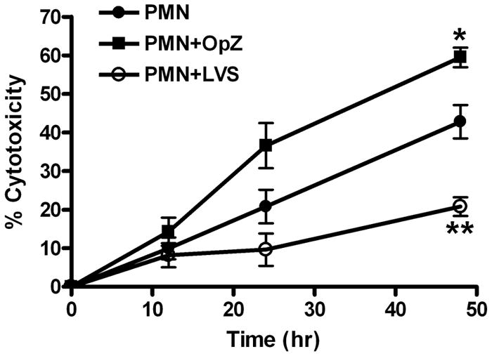
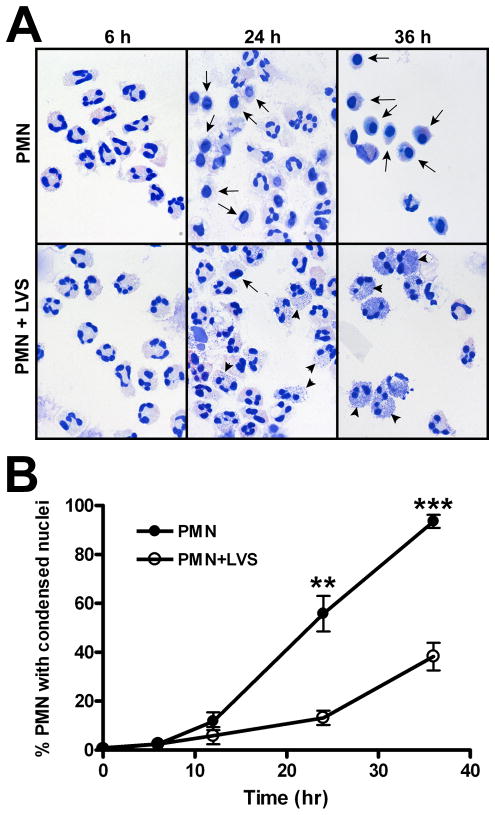
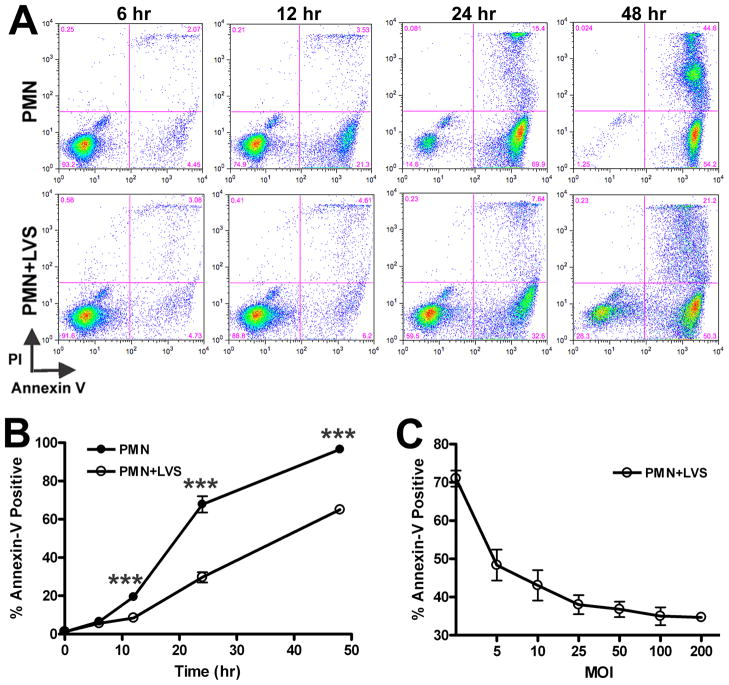
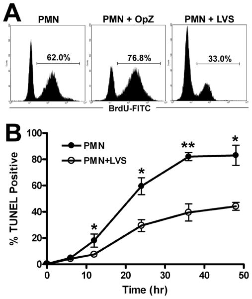
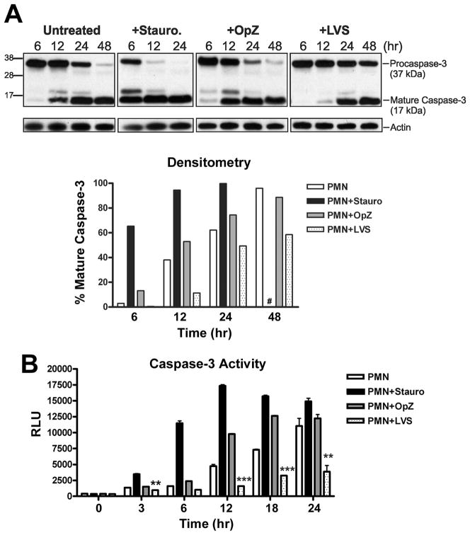
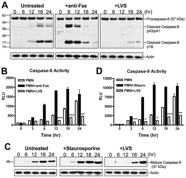
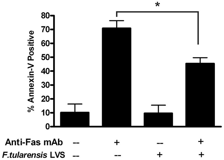
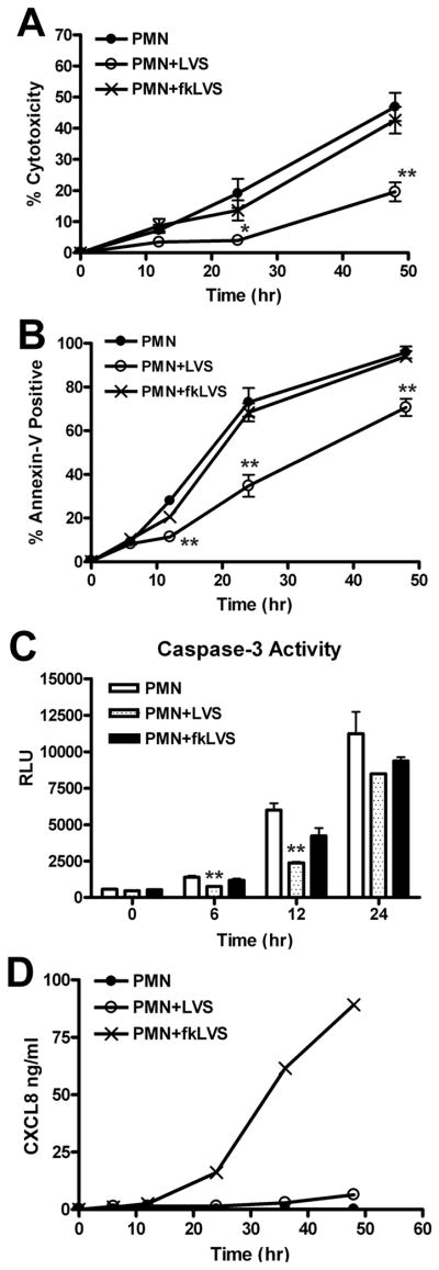
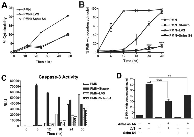
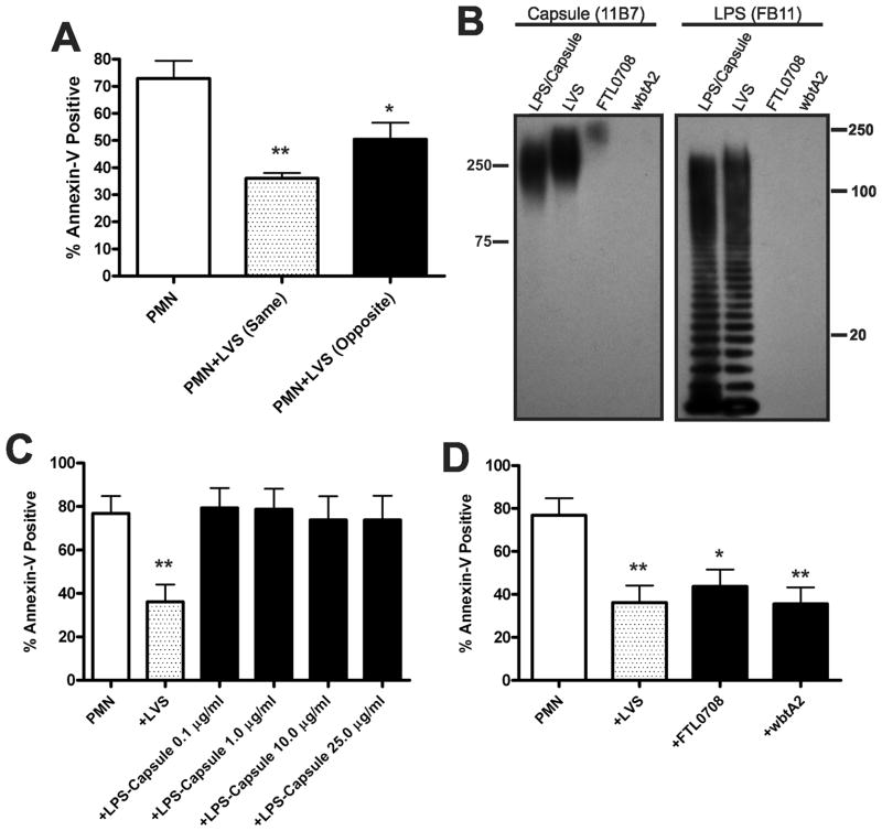
Francisella tularensis is a facultative intracellular bacterium that infects many cell types, including neutrophils. We demonstrated previously that F. tularensis inhibits NADPH oxidase assembly and activity and then escapes the phagosome to the cytosol, but effects on other aspects of neutrophil function are unknown. Neutrophils are short-lived cells that undergo constitutive apoptosis, and phagocytosis typically accelerates this process. We now demonstrate that F. tularensis significantly inhibited neutrophil apoptosis as indicated by morphologic analysis as well as annexin V and TUNEL staining. Thus, ∼80% of infected neutrophils remained viable at 48 h compared with ∼50% of control cells, and ∼40% of neutrophils that ingested opsonized zymosan. In keeping with this finding, processing and activation of procaspases-8, -9, and -3 were markedly diminished and delayed. F. tularensis also significantly impaired apoptosis triggered by Fas crosslinking. Of note, these effects were dose dependent and could be conferred by either intracellular or extracellular live bacteria, but not by formalin-killed organisms or isolated LPS and capsule, and were not affected by disruption of wbtA2 or FTT1236/FTL0708-genes required for LPS O-antigen and capsule biosynthesis. In summary, we demonstrate that F. tularensis profoundly impairs constitutive neutrophil apoptosis via effects on the intrinsic and extrinsic pathways, and thereby define a new aspect of innate immune evasion by this organism. As defects in neutrophil turnover prevent resolution of inflammation, our findings also suggest a mechanism that may in part account for the neutrophil accumulation, granuloma formation, and severe tissue damage that characterizes lethal pneumonic tularemia.


Conflict of interest statement

Conflicts of Interest: None of the authors has a conflict of interest.

Figures

FIGURE 1
FIGURE 1
F. tularensis LVS infects human neutrophils in serum-free media. A, Representative confocal images of PMNs incubated with LVS at a MOI 200:1 for 6, 12, 24, 36 or 48 h at 37°C. Bacteria are shown in red and PMNs were detected by DIC optics and DAPI-staining of nuclear DNA (blue). Arrows indicate infected cells. B, Infection efficiency was quantified by confocal microscopy. Data indicate the percentage of PMNs containing 0, 1–5, 6–10, 11–20, or >20 bacteria/cell at each time point and are the mean ± SEM (n=3). C, Total, extracellular (media), and cell-associated (PMN pellet) LVS were quantified by CFU measurement. Data are the mean ± SEM (n=3). D, Intracellular growth. PMNs were infected with LVS at MOI 200:1 for 12 h, washed extensively to remove extracellular bacteria, and returned to 37°C in sterile media. At the indicated time points, PMNs were lysed with saponin and viable intracellular bacteria were quantified by enumeration of CFU. Data are the mean ± SEM (n=3).
FIGURE 2
FIGURE 2
F. tularensis LVS prolongs neutrophil viability. PMNs were left untreated or were mixed with opsonized zymosan (OpZ; MOI 5:1) or LVS (MOI 200:1) at 37°C. Loss of neutrophil plasma membrane integrity was quantified at 0, 12, 24, and 48 h by measuring release of cytosolic lactate dehydrogenase into the culture medium. Data are the mean ± SEM from three or more independent experiments. * P <0.05 and ** P <0.01 vs. PMNs alone.
FIGURE 3
FIGURE 3
F. tularensis LVS delays the progression of human neutrophils to an apoptotic morphology. Untreated PMNs or cells infected with LVS (MOI 200:1) for the indicated times were fixed and stained using HEMA 3 reagents, and nuclear morphology was analyzed by light microscopy. A, Representative images of untreated and LVS-infected PMNs. Arrows at 24 and 36 h indicate PMNs with condensed, apoptotic nuclei. Arrowheads indicate intracellular LVS that also stain with HEMA 3. B, Pooled data indicate the percentage of cells with apoptotic nuclei determined by microscopic evaluation of >300 cells per time point and are the mean ± SEM (n=4). ** P <0.01 and *** P <0.001.
FIGURE 4
FIGURE 4
F. tularensis LVS reduces Annexin V staining of neutrophils in a dose-dependent manner. Uninfected cells or PMNs infected with LVS were double-stained with Annexin V-FITC and propidium iodide (PI) and then analyzed by flow cytometry. A, Representative dot plots of control PMNs and cells infected with LVS (MOI 200:1) at 6, 12, 24 and 48 h. The percentage of cells in each quadrant is indicated. B, Pooled flow cytometry data indicate the fraction of Annexin-V-positive PMNs at the indicated time points. Data are the mean ± SEM (n=12). *** P <0.001 for control vs. LVS-infected PMNs. Where not visible, error bars are smaller than symbols. C, Neutrophils were infected with increasing doses of LVS for 24 h and the Annexin V-positive cells were quantified by flow cytometry. Data are the mean ± SEM (n=3).
FIGURE 5
FIGURE 5
F. tularensis LVS inhibits neutrophil DNA fragmentation. PMNs were left untreated or incubated with opsonized zymosan (OpZ, MOI 5:1) or LVS (MOI 200:1). Nuclear DNA fragmentation was detected using a modified TUNEL assay and flow cytometry. A, Representative histograms obtained for control, OpZ or LVS treated PMNs after 24 h at 37°C. B, Pooled data indicate the percentage of TUNEL-positive cells and are the mean ± SEM (n≥3). *P <0.05 and ** P <0.01 for control vs. LVS-infected PMNs.
FIGURE 6
FIGURE 6
Procaspase-3 processing and caspase-3 activity are impaired by LVS. PMNs were left untreated or were incubated with staurosporine (stauro., 1 μM), opsonized zymosan (OpZ, MOI 5:1), or LVS (MOI 200:1). A, At the indicated time points, procaspase-3 processing was detected by Western blotting of cell lysates. The caspase-3 mAb recognizes both procaspase-3 (37 kDa) and the large subunit of active caspase-3 (17 kDa). Actin was used as a loading control. Data shown are from one experiment representative of four. Lower panel shows the percentage of mature caspase-3 in each sample determined by densitometric analysis of each immunoblot normalized to the actin loading control. #, 48 h time point not determined for staurosporine-treated PMNs. B, Caspase-3 activity was assessed using a caspase-3-specific proluminogenic substrate. Data indicate relative luminescence units (RLU) and are the mean ± SEM of triplicate samples from one representative experiment (n≥3). ** P <0.01 and *** P <0.001 for LVS-infected vs. control PMN.
FIGURE 7
FIGURE 7
LVS inhibits processing and activation of the initiator caspases -8 and -9. Untreated PMNs or cells treated with staurosporine (1 μM), anti-Fas IgM (500 ng/ml), or LVS (MOI 200:1) were incubated at 37°C. A, Immunoblots of cell lysates obtained at the indicated time points show full length procaspase-8 (57 kDa), cleavage intermediates p43/p41, and mature caspase-8 (p18). Actin immunoblots demonstrate equal loading. Data shown are from one experiment representative of two. B, Caspase-8 activity was assessed using a caspase-8-specific proluminogenic substrate. Data indicate relative luminescence units (RLU) and are the mean ± SEM of triplicate samples from a representative experiment (n≥3). ** P <0.01 and *** P <0.001 for LVS-infected vs. control PMNs. C, Mature caspase-9 was detected in PMN lysates using an antibody specific for the 37 kDa processed enzyme. Actin served as a loading control. Data shown are representative of two independent experiments. D, Caspase-9 activity was measured using a caspase-9-specific proluminogenic substrate. Data indicate relative luminescence unit (RLU) and are the mean ± SEM of triplicate samples from one representative experiment (n≥3). *P <0.05, ** P <0.01 and *** P <0.001 for LVS-treated vs. control PMNs as indicated.
FIGURE 8
FIGURE 8
F. tularensis LVS inhibits Fas-induced apoptosis. Neutrophils were preincubated with LVS (MOI 200:1) for 1 h, and then treated with 500 ng/ml anti-Fas IgM for an additional 6 h. Apoptosis was measured using Annexin V staining and flow cytometry. Data indicate the percentage of Annexin V-positive cells and are the mean ± SEM (n=3).* P <0.05.
FIGURE 9
FIGURE 9
Formalin-killed LVS does not delay neutrophil apoptosis. PMNs were left untreated or were infected with live or formalin-killed LVS (fkLVS) at MOI 200:1. A, PMN lysis was quantified by measuring LDH activity in cell-free supernatants. Data indicate percent cytotoxicity and are the mean ± SEM (n=3). B, PMN apoptosis was quantified using Annexin V staining and flow cytometry. Data are the mean ± SEM (n=3). C, Caspase-3 activity was assessed using a specific luminescence assay. RLU data are the mean ± SEM of triplicate samples from one experiment representative of two. For panels A–C, statistically significant differences between control and LVS-infected PMNs are * P <0.05 and ** P <0.01. D, CXCL8 in media of control, LVS- or fkLVS-infected PMNs was quantified by ELISA. Data shown are from one experiment representative of three.
FIGURE 10
FIGURE 10
Virulent F. tularensis stain Schu S4 impairs spontaneous and Fas-triggered apoptosis. Neutrophils were left untreated, infected with Schu S4 or LVS (at MOI 200:1), or treated with staurosporine (1 μM) as indicated. A, PMN death was quantified by measurement of LDH release. Data shown are from one experiment representative of three. B, Data indicate the percentage of cells with apoptotic nuclei determined by microscopic evaluation of >300 cells per time point and are the mean ± SEM (n=2). *** P <0.001 vs. untreated control. C, Caspase-3 activity was assessed as described above. RLU data are the mean ± SEM of triplicate samples from a representative experiment. *** P <0.001 vs. untreated controls. D, Like LVS, Schu S4 inhibits apoptosis triggered by Fas crosslinking. PMNs were preincubated with Schu S4 or LVS (MOI 200:1) for 1 h, and then treated with 500 ng/ml anti-Fas IgM for an additional 6 h. Apoptosis was assessed by analysis of nuclear morphology after HEMA-3 staining. Data indicate the percentage of cells with condensed nuclei and are the mean ± SEM (n=2). ** P <0.01 and ***P<0.001 as indicated.
FIGURE 11
FIGURE 11
Delayed apoptosis does not require bacterial binding to PMNs, capsular polysaccharides, or LPS O-antigen. A, Delayed apoptosis does not require LVS binding. PMNs were added to the lower chamber of a Transwell containing a 0.4 μm filter support. Where noted, LVS was added to the lower (same) or upper (opposite) chamber of the Transwell at MOI 200:1. Apoptosis was quantified after 24 h at 37°C. Data indicate the percentage of Annexin V-positive PMNs and are the mean ± SEM (n=3). * P <0.05 and ** P <0.01 vs. PMNs alone. B–D, Major surface polysaccharides of LVS are dispensable for delayed PMN apoptosis. B, Lysates prepared from wild-type LVS or isogenic FTL0708 and wbtA2 mutants were analyzed for the presence of capsule and LPS O-antigen by immunoblotting with mAbs 11B7 and FB11, respectively. An LPS and capsule-enriched preparation from wild-type LVS was used as positive control. C, Apoptosis of control PMNs, cells infected with LVS (MOI 200:1), or cells treated with the indicated amounts of isolated LPS and capsule was assayed after 24 h at 37°C using Annexin V staining and flow cytometry. Data are the mean ± SEM (n=4). ** P <0.01 for LVS-infected vs. control PMNs. D, PMNs were left untreated or were infected with wild-type LVS, FTL0708, or wbtA2 (at MOI 200:1). 24 h later, the percentage of Annexin V-positive cells was determined. Data indicate the mean ± SEM (n=4). * P <0.05 and ** P <0.01 vs. PMNs alone.

Similar articles

Cited by

References

    1. Kennedy AD, DeLeo FR. Neutrophil apoptosis and the resolution of infection. Immunol Res. 2009;43:25–61. - PubMed
    1. Nauseef WM. How human neutrophils kill and degrade microbes: an integrated view. Immunol Rev. 2007;219:88–102. - PubMed
    1. Kobayashi SD, Braughton KR, Whitney AR, Voyich JM, Schwan TG, Musser JM, DeLeo FR. Bacterial pathogens modulate an apoptosis differentiation program in human neutrophils. Proc Natl Acad Sci, USA. 2003;100:10948–10953. - PMC - PubMed
    1. Savill JS, Wyllie AH, Henson JE, Walport MJ, Henson PM, Haslett C. Macrophage phagocytosis of aging neutrophils in inflammation. Programmed cell death in the neutrophil leads to its recognition by macrophages. J Clin Invest. 1989;83:865–875. - PMC - PubMed
    1. Watson RW, Redmond HP, Wang JH, Condron C, Bouchier-Hayes D. Neutrophils undergo apoptosis following ingestion of Escherichia coli. J Immunol. 1996;156:3986–3992. - PubMed

Publication types

MeSH terms