

Serodiagnosis efficacy and immunogenicity of the fusion protein of Mycobacterium tuberculosis composed of the 10-kilodalton culture filtrate protein, ESAT-6, and the extracellular domain fragment of PPE68
- PMID: 22357648
- PMCID: PMC3318282
- DOI: 10.1128/CVI.05708-11
Serodiagnosis efficacy and immunogenicity of the fusion protein of Mycobacterium tuberculosis composed of the 10-kilodalton culture filtrate protein, ESAT-6, and the extracellular domain fragment of PPE68
Abstract
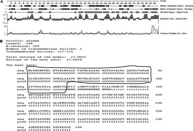
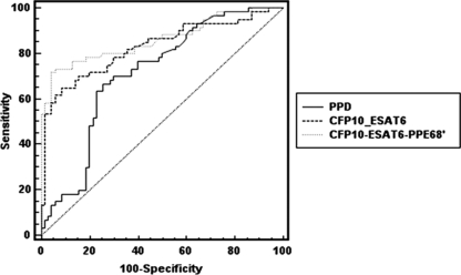
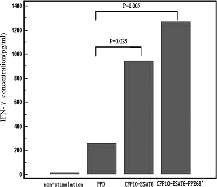
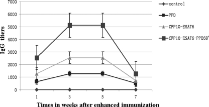
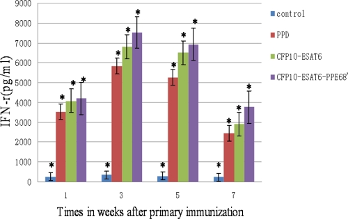
In order to identify immunodominant antigens of Mycobacterium tuberculosis that may be used in the serodiagnosis of active tuberculosis (TB), we designed an M. tuberculosis fusion protein consisting of CFP-10 (10-kDa culture filtrate protein), ESAT-6 (6-kDa early secreted antigenic target), and the extracellular domain fragment of PPE68 (PPE68'). Then, the coding sequences of the three proteins were inserted into a prokaryotic expression vector, pET-32a(+). To enhance the immunological response, the proteins were linked together. The fusion proteins with a 6 × His tag were successfully overexpressed in Escherichia coli BL21 and purified. The purified proteins were applied for detection of the total IgG titer by using an enzyme-linked immunosorbent assay (ELISA) with human sera from well-characterized TB cases and the control cases, and results were compared to those with purified protein derivative tuberculin (PPD). The ELISA results showed that among 140 cases of confirmed active TB and 70 control cases, CFP-10-ESAT-6-PPE68' had a sensitivity of 73.3% and specificity of 94.3%, compared to a sensitivity of 66.7% and specificity of 74.3% for PPD and a sensitivity of 65% and specificity of 91.4% for CFP-10-ESAT-6. In addition, the fusion protein CFP-10-ESAT-6-PPE68' stimulated a higher level of antigen-specific gamma interferon (IFN-γ) release for active-TB patients than PPD and CFP-10-ESAT-6. After immunization of C57BL/6 mice, the findings indicated that the total IgG titers and the concentrations of IFN-γ in mice immunized by CFP-10-ESAT-6-PPE68' were high and induced strong, long-term humoral immunity compared to results with PPD and CFP-10-ESAT-6. Thus, our study indicates that the fusion protein CFP-10-ESAT-6-PPE68' may be useful as an immunodominant antigen for the serodiagnosis of active TB.
Figures
Similar articles
-
Generation of Mycobacterium tuberculosis-specific recombinant antigens and evaluation of the clinical value of antibody detection for serological diagnosis of pulmonary tuberculosis.Int J Mol Med. 2013 Mar;31(3):751-7. doi: 10.3892/ijmm.2013.1254. Epub 2013 Jan 22. Int J Mol Med. 2013. PMID: 23338746
-
Humoral immune responses against the Mycobacterium tuberculosis 38-kilodalton, MTB48, and CFP-10/ESAT-6 antigens in tuberculosis.Clin Vaccine Immunol. 2010 Mar;17(3):372-5. doi: 10.1128/CVI.00287-09. Epub 2010 Jan 6. Clin Vaccine Immunol. 2010. PMID: 20053875 Free PMC article.
-
Cell envelope protein PPE68 contributes to Mycobacterium tuberculosis RD1 immunogenicity independently of a 10-kilodalton culture filtrate protein and ESAT-6.Infect Immun. 2004 Apr;72(4):2170-6. doi: 10.1128/IAI.72.4.2170-2176.2004. Infect Immun. 2004. PMID: 15039340 Free PMC article.
-
[Evolution of IGRA researches].Kekkaku. 2008 Sep;83(9):641-52. Kekkaku. 2008. PMID: 18979999 Review. Japanese.
-
Application of antigenic biomarkers for Mycobacterium tuberculosis.J Zhejiang Univ Sci B. 2020 Nov.;21(11):856-870. doi: 10.1631/jzus.B2000325. J Zhejiang Univ Sci B. 2020. PMID: 33150770 Free PMC article. Review.
Cited by
-
Immunological Characterization of Proteins Expressed by Genes Located in Mycobacterium tuberculosis-Specific Genomic Regions Encoding the ESAT6-like Proteins.Vaccines (Basel). 2021 Jan 7;9(1):27. doi: 10.3390/vaccines9010027. Vaccines (Basel). 2021. PMID: 33430286 Free PMC article. Review.
-
Surface Enhanced CdSe/ZnS QD/SiNP Electrochemical Immunosensor for the Detection of Mycobacterium Tuberculosis by Combination of CFP10-ESAT6 for Better Diagnostic Specificity.Materials (Basel). 2019 Dec 31;13(1):149. doi: 10.3390/ma13010149. Materials (Basel). 2019. PMID: 31906075 Free PMC article.
-
Immunological effects of the PE/PPE family proteins of Mycobacterium tuberculosis and related vaccines.Front Immunol. 2023 Sep 27;14:1255920. doi: 10.3389/fimmu.2023.1255920. eCollection 2023. Front Immunol. 2023. PMID: 37841250 Free PMC article. Review.
-
Genomic comparison between Mycobacterium bovis and Mycobacterium microti and in silico analysis of peptide-based biomarkers for serodiagnosis.Front Vet Sci. 2024 Sep 20;11:1446930. doi: 10.3389/fvets.2024.1446930. eCollection 2024. Front Vet Sci. 2024. PMID: 39372902 Free PMC article.
-
Aqueous extract-mediated green synthesis of CuO nanoparticles: Potential anti-tuberculosis agents.Food Sci Nutr. 2024 May 30;12(8):5907-5921. doi: 10.1002/fsn3.4227. eCollection 2024 Aug. Food Sci Nutr. 2024. PMID: 39139956 Free PMC article.
References
-
- Berthet FX, Rasmussen PB, Rosenkrands I, Andersen P, Gicquel B. 1998. A Mycobacterium tuberculosis operon encoding ESAT-6 and a novel low-molecular-mass culture filtrate protein (CFP-10). Microbiology 144:3195–3203 - PubMed
-
- Brusasca PN, et al. 2001. Immunological characterization of antigens encoded by the RD1 region of the Mycobacterium tuberculosis genome. Scand. J. Immunol. 54:448–452 - PubMed
Publication types
MeSH terms
Substances
LinkOut - more resources
Full Text Sources
Other Literature Sources
Medical
Miscellaneous
