. 2012 May;81(5):748-58.
doi: 10.1124/mol.112.077586. Epub 2012 Feb 22.

Lapatinib and obatoclax kill tumor cells through blockade of ERBB1/3/4 and through inhibition of BCL-XL and MCL-1

Affiliations

Lapatinib and obatoclax kill tumor cells through blockade of ERBB1/3/4 and through inhibition of BCL-XL and MCL-1

Nichola Cruickshanks et al. Mol Pharmacol. 2012 May.

Abstract

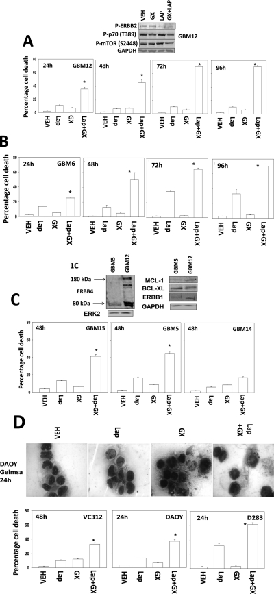
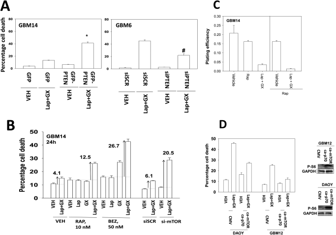
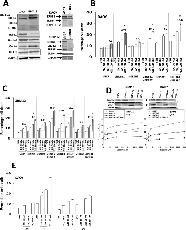
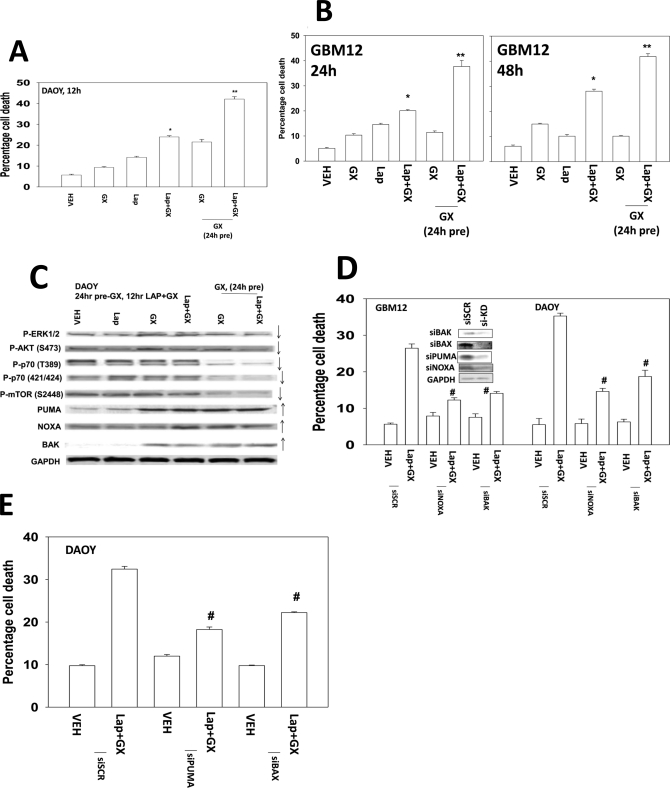
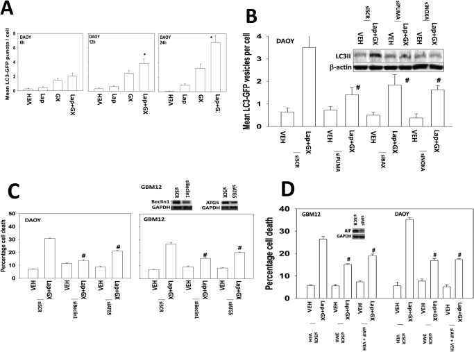
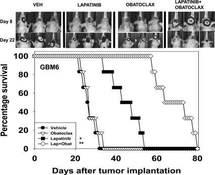
Prior studies in breast cancer cells have shown that lapatinib and obatoclax interact in a greater than additive fashion to cause cell death and do so through a toxic form of autophagy. The present studies sought to extend our analyses to the central nervous system (CNS) tumor cells and to further define mechanisms of drug action. Lapatinib and obatoclax killed multiple CNS tumor isolates. Cells lacking PTEN (phosphatase and tensin homolog on chromosome 10) function were relatively resistant to drug combination lethality; expression of PTEN in PTEN-null cells restored drug sensitivity, and knockdown of PTEN promoted drug resistance. On the basis of knockdown of ERBB1-4 (erythroblastic leukemia viral oncogene homolog 1-4), we discovered that the inhibition of ERBB1/3/4 receptors were most important for enhancing obatoclax lethality rather than ERBB2. In parallel, we noted in CNS tumor cells that knockdown of BCL-xL (B-cell lymphoma-extra large)and MCL-1 (myeloid cell leukemia-1) interacted in an additive fashion to facilitate lapatinib lethality. Pretreatment of tumor cells with obatoclax enhanced the lethality of lapatinib to a greater extent than concomitant treatment. Treatment of animals carrying orthotopic CNS tumor isolates with lapatinib- and obatoclax-prolonged survival. Altogether, our data show that lapatinib and obatoclax therapy could be of use in the treatment of tumors located in the CNS.


Figures

Fig. 1.
Fig. 1.
Lapatinib and obatoclax interact to kill multiple CNS tumor cells but not those cells lacking PTEN function/expression. A, GBM12 cells were treated with vehicle (VEH, DMSO), lapatinib (lap, 1 μM), and/or obatoclax (GX, 50 nM) as indicated. Cells were isolated 24 to 96 h after exposure, and viability was determined by trypan blue exclusion (n = 3, ±S.E.M.).*, P < 0.05 greater than vehicle control. Top blot, drug combination causes inactivation of ERBB2, p70 S6K, and mTOR. B, GBM6 cells were treated with vehicle (DMSO), lapatinib (lap, 1 μM), and/or obatoclax (GX, 50 nM) as indicated. Cells were isolated 24 to 96 h after exposure, and viability was determined by trypan blue exclusion (n = 3, ±S.E.M.).*, P < 0.05 greater than vehicle control. C, GBM5, GBM14, and GBM15 cells were treated with vehicle (DMSO), lapatinib (1 μM), and/or obatoclax (GX, 50 nM) as indicated. Cells were isolated 48 h after exposure, and viability was determined by trypan blue exclusion (n = 3, ± S.E.M.).*, P < 0.05 greater than vehicle control. Top blot, the expression of ERBB1, ERBB4, MCL-1, and BCL-xL in GBM5 and GBM12 cells. D, VC312, DAOY, and D283 pediatric CNS tumor cells were treated with vehicle (DMSO), lapatinib (1 μM), and/or obatoclax (GX, 50 nM) as indicated. Cells were isolated 24 to 48 h after exposure, and viability was determined by trypan blue exclusion (n = 3, ±S.E.M.).*, P < 0.05 greater than vehicle control. Top, Giemsa staining of DAOY cells 24 h after drug treatment.
Fig. 2.
Fig. 2.
Loss of PTEN function renders tumor cells resistant to lapatinib and obatoclax treatment. A, GBM14 cells were transfected with either control (GFP) or with a plasmid to express PTEN (GFP-PTEN). GBM6 cells were transfected with either a control plasmid short hairpin RNA or a plasmid to express a short hairpin RNA to knock down PTEN. Twenty-four hours after transfection, cells were treated with vehicle (VEH, DMSO) or with lapatinib (lap, 1 μM) and obatoclax (GX, 50 nM). Cells were isolated 24 h after exposure, and viability was determined by trypan blue exclusion (n = 3, ±S.E.M.). B, GBM14 cells were treated with vehicle (DMSO) or with lapatinib (1 μM) and obatoclax (GX, 50 nM) in the presence or absence of rapamycin (Rap, 10 nM) or BEZ-235 (BEZ, 50 nM). Parallel sets of cells were transfected to knock down mTOR. Cells were isolated 24 after exposure, and viability was determined by trypan blue exclusion (n = 3, ±S.E.M.). C, GBM14 cells were plated as single cells in sextuplicate and were treated for 48 h with vehicle, lapatinib (1 μM) + obatoclax (GX, 50 nM), rapamycin (10 nM), or the drug combination as indicated. Colony formation 14 days later was determined (n = 3, ±S.E.M.). D, DAOY and GBM12 cells were transfected with empty vector [cytomegalovirus (CMV)] or with plasmids to express activated p70 S6K and activated mTOR. Cells were treated with vehicle (DMSO) or with lapatinib (1 μM) and obatoclax (GX, 50 nM). Cells were isolated 24 h after exposure, and viability was determined by trypan blue exclusion (n = 3, ± S.E.M.).
Fig. 3.
Fig. 3.
Dissecting the roles of ERBB receptors and protective BCL-2 family members in the toxic interaction between lapatinib and obatoclax. A, expression of ERBB1-4, BCL-xL, and MCL-1 in CNS tumor cells and the knock down of these proteins by siRNA. DAOY (B) and GBM12 (C) cells had expression of the indicated receptors knocked down. Cells were treated with vehicle (DMSO) or with obatoclax (GX, 10–100 nM). Cells were isolated 24 h after exposure, and viability was determined by trypan blue exclusion (n = 3, ± S.E.M.); the values shown indicate the true percentage of cell death above its matched vehicle control:*, P < 0.05 greater than vehicle control; **, P < 0.05 greater than the same value under parallel other conditions. D, DAOY and GBM12 cells had expression of the indicated protective BCL-2 family proteins (MCL-1, BCL-xL) knocked down. Cells were treated with vehicle (DMSO) or with lapatinib (100–3000 nM). Cells were isolated 24 h after exposure, and viability was determined by trypan blue exclusion (n = 3, ±S.E.M.); the values shown indicate the true percentage of cell death above its matched vehicle control. E, DAOY cells were treated with 1 μM lapatinib in the presence of obatoclax (GX, 10–100 nM) and ABT-263 (ABT, 10–100 nM). Cells were isolated 24 h after exposure, and viability was determined by trypan blue exclusion (n = 3, ±S.E.M.).*, P < 0.05 greater than vehicle control.
Fig. 4.
Fig. 4.
Pretreatment of cells with obatoclax promotes greater drug combination toxicity than concomitant drug treatment. A, DAOY cells were pretreated with either vehicle (VEH, DMSO) or with obatoclax (GX, 50 nM). After 24 h, as indicated, cells were treated with lapatinib (1 μM) and/or obatoclax (GX, 50 nM). Cells were isolated after 12 h, and viability was determined by trypan blue exclusion (n = 3, ±S.E.M.)*, P < 0.05 greater than vehicle control; **, P < 0.05 greater than parallel value in nonpretreated cells. B, GBM12 cells were pretreated with either vehicle (DMSO) or with obatoclax (50 nM). After 24 h, as indicated, cells were treated with lapatinib (1 μM) and/or obatoclax (GX, 50 nM). Cells were isolated after 24 to 48 h, and viability was determined by trypan blue exclusion (n = 3, ± S.E.M.).*, P < 0.05 greater than vehicle control; **, P < 0.05 greater than parallel value in nonpretreated cells. C, DAOY cells (see A) were isolated, and immunoblotting performed on cell lysates. D and E, DAOY and GBM12 cells were transfected to knock down PUMA and/or NOXA expression or to knock down BAK or BAX expression. Twenty-four hours later, cells were treated with vehicle (DMSO) or with lapatinib (1 μM) and obatoclax (GX, 50 nM). Cells were isolated 24 h later, and viability was determined by trypan blue exclusion (n = 3, ±S.E.M.). #, P < 0.05 less than vehicle control value.
Fig. 5.
Fig. 5.
Lapatinib and obatoclax interact to kill CNS tumor cells through a toxic form of autophagy. A, DAOY cells were transfected with a plasmid to express LC3-GFP. Cells were treated with lapatinib (1 μM) and/or obatoclax (GX, 50 nM), and the number of LC3-GFP punctae was determined over 6 to 24 h (n = 3, ±S.E.M.)*, P < 0.05 greater than vehicle control. B, DAOY cells were transfected to knock down PUMA and/or NOXA expression or to knock down BAX expression in parallel with a plasmid to express LC3-GFP. Cells were treated with lapatinib (1 μM) and/or obatoclax (GX, 50 nM), and the number of LC3-GFP punctae was determined over 24 h (n = 3, ±S.E.M.). #, P < 0.05 less than vehicle control. C, DAOY and GBM12 cells were transfected with a scrambled siRNA (siSCR) or siRNA molecules to knock down Beclin-1 or ATG5. Twenty-four hours after transfection, cells were treated with vehicle (DMSO) or with lapatinib (1 μM) and obatoclax (GX, 50 nM). Cells were isolated 24 h later, and viability was determined by trypan blue exclusion (n = 3, ± S.E.M.). #, P < 0.05 less than vehicle control. D, DAOY and GBM12 cells were transfected with a scrambled siRNA (siSCR) or a siRNA molecule to knock down apoptosis-inducing factor (AIF). Twenty-four hours after transfection, cells were treated vehicle (PBS) followed as indicated by 3-methyl adenine (3MA, 5 mM) followed by with vehicle (DMSO) or with lapatinib (1 μM) and obatoclax (GX, 50 nM). Cells were isolated 24 h later, and viability was determined by trypan blue exclusion (n = 3, ± S.E.M.). #, P < 0.05 less than vehicle control.
Fig. 6.
Fig. 6.
Lapatinib and obatoclax prolong survival of animals with intracranial tumors. GBM6-luc cells (106) were implanted into the brains of mice. Eight days after implantation animals were treated with vehicle (VEH, Cremophor EL), lapatinib (100 mg/kg b.i.d.), obatoclax (5 mg/kg q.i.d.), or the drug combination for 5 days. Top, animals carrying tumors show regression 14 days after initiation of drug combination exposure as judged by bioluminescent imaging. Bottom, animals exposed to lapatinib show elevated survival compared with vehicle control (two studies, n = 6, ±S.E.M.).*, P < 0.05 greater than vehicle treatment; **, P < 0.05 exposure to lapatinib and obatoclax causes greater survival than lapatinib alone.

Similar articles

Cited by

References

    1. Alva AS, Gultekin SH, Baehrecke EH. (2004) Autophagy in human tumors: cell survival or death? Cell Death Differ 11:1046–1048 - PubMed
    1. Awada A, Saliba W, Bozovic-Spasojevic I. (2011) Lapatinib ditosylate: expanding therapeutic options for receptor tyrosine-protein kinase erbB-2-positive breast cancer. Drugs Today (Barc) 47:335–345 - PubMed
    1. Berezowska S, Schlegel J. (2011) Targeting ErbB receptors in high-grade glioma. Curr Pharm Des 17:2468–2487 - PubMed
    1. Bigner SH, Vogelstein B. (1990) Cytogenetics and molecular genetics of malignant gliomas and medulloblastoma. Brain Pathol 1:12–18 - PubMed
    1. Depowski PL, Rosenthal SI, Ross JS. (2001) Loss of expression of the PTEN gene protein product is associated with poor outcome in breast cancer. Mod Pathol 14:672–676 - PubMed

Publication types

MeSH terms