. 2012 May;107(10):2901-11.
doi: 10.1152/jn.00850.2011. Epub 2012 Feb 22.

Impaired crossed facilitation of the corticospinal pathway after cervical spinal cord injury

Affiliations

Impaired crossed facilitation of the corticospinal pathway after cervical spinal cord injury

Karen L Bunday et al. J Neurophysiol. 2012 May.

Abstract

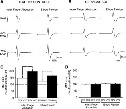
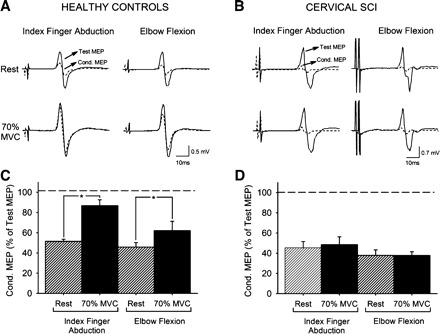
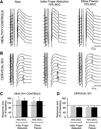
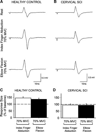
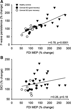
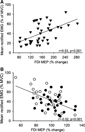
In uninjured humans, it is well established that voluntary contraction of muscles on one side of the body can facilitate transmission in the contralateral corticospinal pathway. This crossed facilitatory effect may favor interlimb coordination and motor performance. Whether this aspect of corticospinal function is preserved after chronic spinal cord injury (SCI) is unknown. Here, using transcranial magnetic stimulation, we show in patients with chronic cervical SCI (C(5)-C(8)) that the size of motor evoked potentials (MEPs) in a resting intrinsic hand muscle remained unchanged during increasing levels of voluntary contraction with a contralateral distal or proximal arm muscle. In contrast, MEP size in a resting hand muscle was increased during the same motor tasks in healthy control subjects. The magnitude of voluntary electromyography was negatively correlated with MEP size after chronic cervical SCI and positively correlated in healthy control subjects. To examine the mechanisms contributing to MEP crossed facilitation we examined short-interval intracortical inhibition (SICI), interhemispheric inhibition (IHI), and motoneuronal behavior by testing F waves and cervicomedullary MEPs (CMEPs). During strong voluntary contractions SICI was unchanged after cervical SCI and decreased in healthy control subjects compared with rest. F-wave amplitude and persistence and CMEP size remained unchanged after cervical SCI and increased in healthy control subjects compared with rest. In addition, during strong voluntary contractions IHI was unchanged in cervical SCI compared with rest. Our results indicate that GABAergic intracortical circuits, interhemispheric glutamatergic projections between motor cortices, and excitability of index finger motoneurons are neural mechanisms underlying, at least in part, the lack of crossed corticospinal facilitation observed after SCI. Our data point to the spinal motoneurons as a critical site for modulating corticospinal transmission after chronic cervical SCI.


Figures

Fig. 1.
Fig. 1.
Motor evoked potentials (MEPs). A and B: MEPs recorded from the resting first dorsal interosseous (FDI) of a representative healthy control subject (A) and in a patient with cervical spinal cord injury (SCI) (B) while the other side remained at rest or performed 30% or 70% maximal voluntary contraction (MVC) during index finger abduction or elbow flexion. C and D: group data for healthy control subjects (n = 10, C) and cervical SCI patients (n = 14, D). The x-axis shows the MVC levels tested. The y-axis shows the size of the FDI MEP as % of the baseline FDI MEP. Note the increase in FDI MEP size during contralateral index finger abduction and elbow flexion in healthy control subjects but not in patients with cervical SCI. Error bars indicate SE. *P < 0.05.
Fig. 2.
Fig. 2.
Short-interval intracortical inhibition (SICI). A and B: SICI recorded from the resting FDI of a representative healthy control (A) and a patient with cervical SCI (B). The test MEP (solid traces) and conditioned MEP (Cond. MEP, dashed traces) are indicated by arrows. C and D: group data for healthy control subjects (n = 8, C) and cervical SCI patients (n = 8, D). The x-axis shows all conditions tested. The y-axis shows the magnitude of the conditioned MEP expressed as % of the test MEP. The horizontal dashed line represents the size of the test MEP. Note that SICI decreased during index finger abduction and elbow flexion in healthy control subjects but not after cervical SCI. Error bars indicate SE. *P < 0.05.
Fig. 3.
Fig. 3.
F waves. A and B: M waves and F waves recorded from the resting FDI of a representative healthy control subject (A) and a patient with cervical SCI (B) during index finger abduction and elbow flexion. C and D: group data for healthy control subjects (n = 8, C) and cervical SCI patients (n = 9, D). The x-axis shows all conditions tested [F-wave persistence (gray bars) and F-wave mean amplitude (black bars) during 70% of MVC of index finger abduction and elbow flexion]. The y-axis shows F-wave persistence (% of F waves present on each set) and F-wave amplitude [% of maximal motor response (M-max)]. Note that F-wave persistence and mean amplitude increased during index finger abduction and elbow flexion in healthy control subjects but not after cervical SCI. Error bars indicate SE. *P < 0.05.
Fig. 4.
Fig. 4.
Cervicomedullary motor evoked potentials (CMEPs). A and B: CMEPs recorded from the resting FDI of a healthy control subject (A) and a patient with cervical SCI (B) while the other side remained at rest or performed 70% of MVC during index finger abduction or elbow flexion. C and D: x-axis shows the conditions tested (70% of MVC during index finger abduction, white bars; 70% of MVC during elbow flexion, black bars), and y-axis shows the size of the FDI CMEP as % of the baseline FDI CMEP. Note the increase in FDI MEP size during contralateral index finger abduction and elbow flexion in the healthy control subject (C) but not in the patient with cervical SCI (D). Error bars indicate SE.
Fig. 5.
Fig. 5.
Correlation analysis between the changes in FDI MEP size during 70% of MVC during the index finger abduction and elbow flexion motor tasks and F-wave persistence (A) and SICI (B) in all subjects tested. In both graphs, the x-axis shows the size of the FDI MEP during 70% of MVC expressed as % of the FDI MEP size at rest. The y-axis shows the F-wave persistence and SICI during 70% of MVC expressed as % of the same measurements taken at rest. Note that larger changes in FDI MEP size were associated with larger F-wave persistence but not with changes in SICI.
Fig. 6.
Fig. 6.
Correlation analysis between the changes in FDI MEP size during 30% and 70% of MVC during the index finger abduction and elbow flexion motor tasks and mean rectified EMG activity exerted during the same motor tasks in healthy control subjects (A, ▾) and patients with cervical SCI with poor (B, ○) and good (B, ●) recovery. In both graphs, x-axis shows the size of the FDI MEP during 30% and 70% of MVC expressed as % of the FDI MEP size at rest and y-axis shows the mean rectified EMG activity in the FDI expressed as % of the MVC. Note that larger changes in mean rectified EMG activity in the FDI muscle were associated with larger FDI MEP size in healthy control subjects and smaller FDI MEP size in patients with SCI.

References

    1. Aimonetti JM, Nielsen JB. Changes in intracortical excitability induced by stimulation of wrist afferents in man. J Physiol 534: 891–902, 2001 - PMC - PubMed
    1. Barthélemy D, Willerslev-Olsen M, Lundell H, Conway BA, Knudsen H, Biering-Sørensen F, Nielsen JB. Impaired transmission in the corticospinal tract and gait disability in spinal cord injured persons. J Neurophysiol 104: 1167–1176, 2010 - PubMed
    1. Bennett DJ, Sanelli L, Cooke CL, Harvey PJ, Gorassini MA. Spastic long-lasting reflexes in the awake rat after sacral spinal cord injury. J Neurophysiol 91: 2247–2258, 2004 - PubMed
    1. Brus-Ramer M, Carmel JB, Martin JH. Motor cortex bilateral motor representation depends on subcortical and interhemispheric interactions. J Neurosci 29: 6196–6206, 2009 - PMC - PubMed
    1. Butler JE, Thomas CK. Effects of sustained stimulation on the excitability of motoneurons innervating paralyzed and control muscles. J Appl Physiol 94: 567–575, 2003 - PubMed

Publication types