

Neuronal projections from V1 to V2 in amblyopia
- PMID: 22357849
- PMCID: PMC3325646
- DOI: 10.1523/JNEUROSCI.4799-11.2012
Neuronal projections from V1 to V2 in amblyopia
Abstract
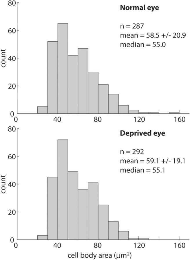
The mechanism of amblyopia in children with congenital cataract is not understood fully, but studies in macaques have shown that geniculate synapses are lost in striate cortex (V1). To search for other projection abnormalities in amblyopia, the pathway from V1 to V2 was examined using a triple-label technique in three animals raised with monocular suture. [(3)H]proline was injected into one eye to label the ocular dominance columns. Cholera toxin B subunit conjugated to gold (CTB-Au) was injected into V2 to label V1 projection neurons. Alternate sections were processed for cytochrome oxidase (CO) and CTB-Au, or dipped for autoradiography. Eight fields of CTB-Au-labeled cells in V1 opposite injection sites were plotted in layers 2/3 or 4B. After thin stripe injection, labeled cells were concentrated in CO patches. Despite column shrinkage, cells in deprived and normal columns were equal in size and density in both layers 2/3 and 4B. After pale or thick stripe injection, labeled cells were concentrated in interpatches. Only 23% of projection neurons originated from deprived columns. This reduction exceeded the degree of column shrinkage, a result explained by the fact that column shrinkage causes disproportionate loss of interpatch territory. These data indicate that early monocular form deprivation does not alter the segregation of patch and interpatch pathways to V2 stripes or cause selective loss or atrophy of V1 projection neurons. The effect of shrinkage of geniculocortical afferents in layer 4C following visual deprivation is not amplified further by attenuation of the amblyopic eye's projections from V1 to V2.
Figures


Similar articles
-
Timing of the critical period for plasticity of ocular dominance columns in macaque striate cortex.J Neurosci. 1997 May 15;17(10):3684-709. doi: 10.1523/JNEUROSCI.17-10-03684.1997. J Neurosci. 1997. PMID: 9133391 Free PMC article.
-
Amblyopia induced by anisometropia without shrinkage of ocular dominance columns in human striate cortex.Proc Natl Acad Sci U S A. 1993 Jun 15;90(12):5494-8. doi: 10.1073/pnas.90.12.5494. Proc Natl Acad Sci U S A. 1993. PMID: 8390668 Free PMC article.
-
V1 interpatch projections to v2 thick stripes and pale stripes.J Neurosci. 2010 May 19;30(20):6963-74. doi: 10.1523/JNEUROSCI.5506-09.2010. J Neurosci. 2010. PMID: 20484638 Free PMC article.
-
Ocular integration in the human visual cortex.Can J Ophthalmol. 2006 Oct;41(5):584-93. doi: 10.1016/S0008-4182(06)80027-X. Can J Ophthalmol. 2006. PMID: 17016529 Review.
-
Neural mechanisms of recovery following early visual deprivation.Philos Trans R Soc Lond B Biol Sci. 2009 Feb 12;364(1515):383-98. doi: 10.1098/rstb.2008.0192. Philos Trans R Soc Lond B Biol Sci. 2009. PMID: 18977734 Free PMC article. Review.
Cited by
-
From Basic Visual Science to Neurodevelopmental Disorders: The Voyage of Environmental Enrichment-Like Stimulation.Neural Plast. 2019 May 6;2019:5653180. doi: 10.1155/2019/5653180. eCollection 2019. Neural Plast. 2019. PMID: 31198418 Free PMC article. Review.
-
Covert spatial attention is functionally intact in amblyopic human adults.J Vis. 2016 Dec 1;16(15):30. doi: 10.1167/16.15.30. J Vis. 2016. PMID: 28033433 Free PMC article.
-
Is the Cortical Deficit in Amblyopia Due to Reduced Cortical Magnification, Loss of Neural Resolution, or Neural Disorganization?J Neurosci. 2015 Nov 4;35(44):14740-55. doi: 10.1523/JNEUROSCI.1101-15.2015. J Neurosci. 2015. PMID: 26538646 Free PMC article.
-
Altered functional interactions between neurons in primary visual cortex of macaque monkeys with experimental amblyopia.J Neurophysiol. 2019 Dec 1;122(6):2243-2258. doi: 10.1152/jn.00232.2019. Epub 2019 Sep 25. J Neurophysiol. 2019. PMID: 31553685 Free PMC article.
-
Early monocular defocus disrupts the normal development of receptive-field structure in V2 neurons of macaque monkeys.J Neurosci. 2014 Oct 8;34(41):13840-54. doi: 10.1523/JNEUROSCI.1992-14.2014. J Neurosci. 2014. PMID: 25297110 Free PMC article.
References
-
- Almog Y, Nemet A. The correlation between visual acuity and color vision as an indicator of the cause of visual loss. Am J Ophthalmol. 2010;149:1000–1004. - PubMed
-
- Bradley A, Dahlman C, Switkes E, De Valois K. A comparison of color and luminance discrimination in amblyopia. Invest Ophthalmol Vis Sci. 1986;27:1404–1409. - PubMed
-
- Butz M, Wörgotter F, van Ooyen A. Activity-dependent structural plasticity. Brain Res Rev. 2009;60:287–305. - PubMed
Publication types
MeSH terms
Substances
Grants and funding
LinkOut - more resources
Full Text Sources
Medical