. 2012 Feb 22;32(8):2683-95.
doi: 10.1523/JNEUROSCI.4125-11.2011.

Neurabin scaffolding of adenosine receptor and RGS4 regulates anti-seizure effect of endogenous adenosine

Affiliations

Neurabin scaffolding of adenosine receptor and RGS4 regulates anti-seizure effect of endogenous adenosine

Yunjia Chen et al. J Neurosci. .

Abstract

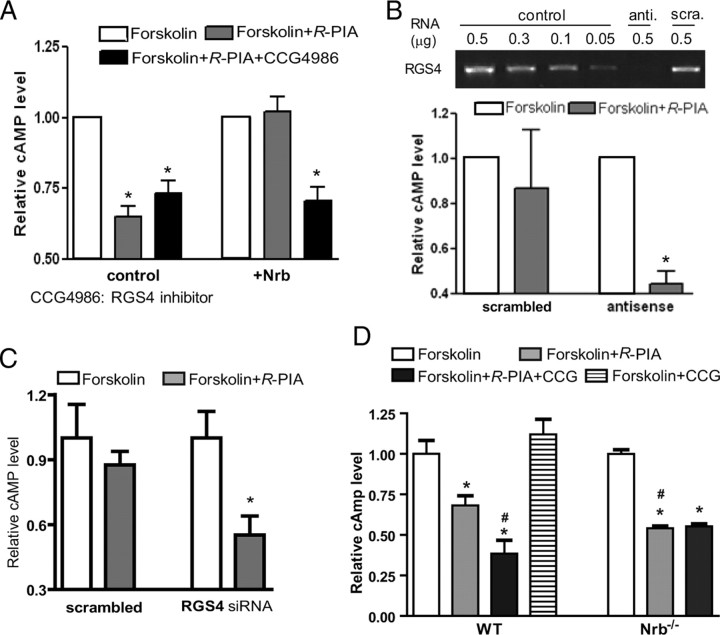
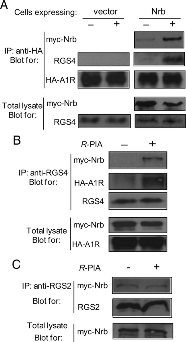
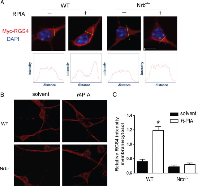
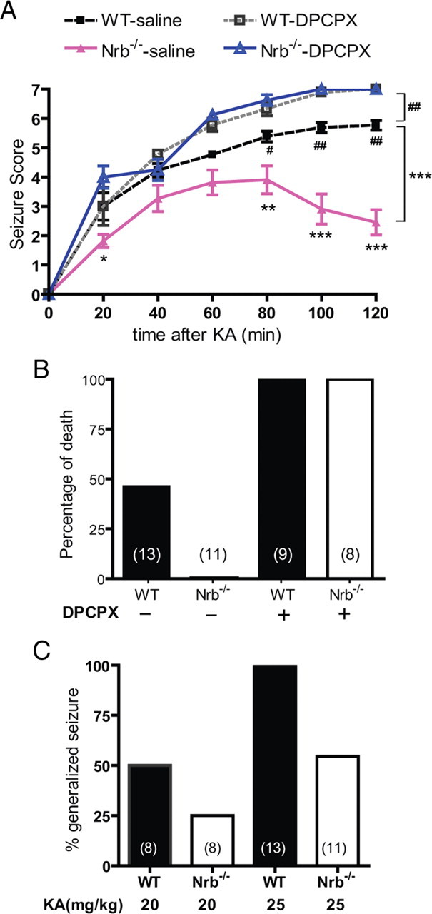
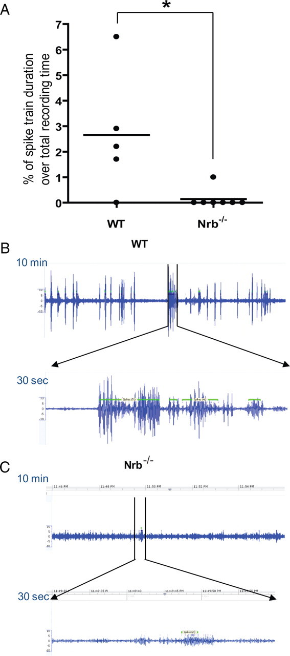
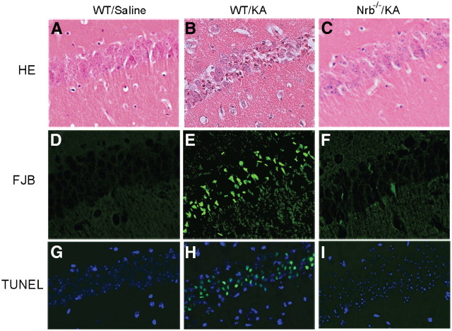
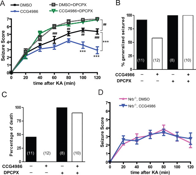
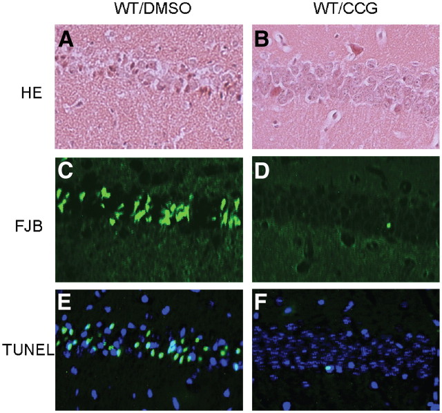
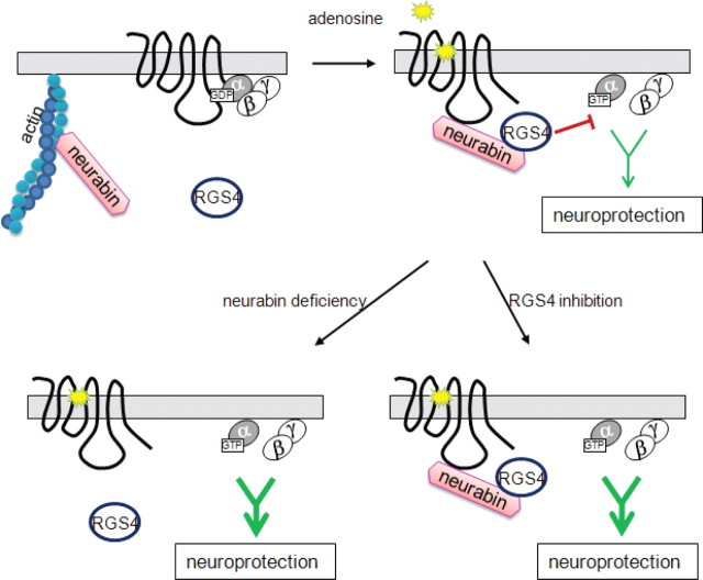
Endogenous adenosine is an essential protective agent against neural damage by various insults to the brain. However, the therapeutic potential of adenosine receptor-directed ligands for neuroprotection is offset by side effects in peripheral tissues and organs. An increase in adenosine receptor responsiveness to endogenous adenosine would enhance neuroprotection while avoiding the confounding effects of exogenous ligands. Here we report novel regulation of adenosine-evoked responses by a neural tissue-specific protein, neurabin. Neurabin attenuated adenosine A(1) receptor (A1R) signaling by assembling a complex between the A1R and the regulator of G-protein signaling 4 (RGS4), a protein known to turn off G-protein signaling. Inactivation of the neurabin gene enhanced A1R signaling and promoted the protective effect of adenosine against excitotoxic seizure and neuronal death in mice. Furthermore, administration of a small molecule inhibitor of RGS4 significantly attenuated seizure severity in mice. Notably, the dose of kainate capable of inducing an ∼50% rate of death in wild-type (WT) mice did not affect neurabin-null mice or WT mice cotreated with an RGS4 inhibitor. The enhanced anti-seizure and neuroprotective effect achieved by disruption of the A1R/neurabin/RGS4 complex is elicited by the on-site and on-demand release of endogenous adenosine, and does not require administration of A1R ligands. These data identify neurabin-RGS4 as a novel tissue-selective regulatory mechanism for fine-tuning adenosine receptor function in the nervous system. Moreover, these findings implicate the A1R/neurabin/RGS4 complex as a valid therapeutic target for specifically manipulating the neuroprotective effects of endogenous adenosine.


Figures

Figure 1.
Figure 1.
The A1R directly interacts with neurabin (Nrb). A, Direct interaction between A1R 3i loop and C-terminal tail (C-tail) and neurabin amino acids 146–453 (Nrb146-453). GST or GST-Nrb146-453 was incubated with in vitro translated [35S]-labeled probe as indicated. B, Neurabin amino acids 331–453 are involved in the direct interaction with the A1R. GST or GST-fused neurabin constructs were incubated with [35S]-labeled probe containing A1R 3i loop and C-tail. C, The 3i loop of A1R is required for a full level of interaction with neurabin. GST or GST-Nrb146-453 was incubated with [35S]-labeled probe containing A1R 3i loop, C-tail, or both, as indicated. Comparable amounts of GST fusion proteins in each reaction were confirmed by Coomassie staining. Free probe represents 1/20 of the input in each reaction.
Figure 2.
Figure 2.
Agonist exposure specifically promotes interaction of the A1R with neurabin (Nrb) but not its homolog spinophilin in intact cells. A, Sequence homology between domains in neurabin and spinophilin. B, Interaction of the A1R with neurabin (left) or spinophilin (right) in cells treated with or without A1R agonist. Cells expressing neurabin alone or coexpressing HA-A1R with Myc-Nrb or Myc-Sp were stimulated with or without 1 μm R-PIA for 5 min, and cell lysates were subjected to immunoisolation assay using an antibody against HA. C, The A1R–neurabin interaction detected with prolonged R-PIA treatment for 30 min. Cells coexpressing HA-A1R and Myc-Nrb were stimulated with 1 μm R-PIA or vehicle for 30 min, and cell lysates were subjected to immunoisolation assay. D, Quantitation of A1R–neurabin interaction representing three to six independent coimmunoisolation experiments. Data are expressed as the fold change of neurabin in complex with the A1R over no stimulation control (defined as onefold). Values are given as the mean ± SEM; *p < 0.05, R-PIA stimulated versus control. E, Endogenous interaction between neurabin and A1R in mouse brain. Mice were given intraperitoneal injections of saline or R-PIA (1 mg/kg), with whole brains isolated and homogenized 30 min postinjection. Detergent-solubilized fractions were then prepared and subjected to coimmunoisolation using equal concentrations of an antibody against either GFP (negative control) or the A1R. Representative blots from three independent experiments are shown. A degradation product of the endogenous neurabin was also detected.
Figure 3.
Figure 3.
Neurabin attenuates A1R-mediated G-protein signaling in cells. A, Neurabin expression attenuates A1R-mediated inhibition of cAMP production. Cells expressing HA-A1R alone or in combination with either neurabin (Nrb) or spinophilin (Sp) were treated with 10 μm forskolin alone or forskolin plus 1 μm R-PIA. Surface receptor density in each experimental group was comparable, as verified by intact cell ELISA using an anti-HA antibody. n = 11 for cells expressing the A1R alone; n = 8 for cells expressing the A1R and neurabin; n = 5 for cells expressing the A1R and spinophilin. Data are expressed as the fold change in cAMP production over forskolin alone control (defined as onefold). *p < 0.05, R-PIA stimulated versus control. B, R-PIA induces similar levels of receptor internalization from the cell surface in cells either with or without exogenous expression of neurabin or spinophilin. Surface receptor density in cells described in A was examined by intact cell ELISA using an anti-HA antibody. *p < 0.05, when comparing cell-surface A1R in R-PIA-treated cells versus nontreated controls.
Figure 4.
Figure 4.
Neurabin attenuates A1R-mediated responses in vivo. A, Neurabin-deficient mice are more sensitive to R-PIA-elicited sedation, as assessed by rotarod latency. The EC50 values for sedation in Nrb−/− (n = 8) and corresponding WT littermates (n = 5) are 1.29 and 2.64 mg/kg, respectively. *p < 0.05; **p < 0.01. B, The α2AR-agonist, UK14,304, induces comparable sedation responses in both WT and Nrb−/− mice as assessed by rotarod latency. n = 5 for both WT and Nrb−/− mice. Error bars indicate mean ± SEM. C, The density of A1Rs is indistinguishable in brain membrane preparations obtained from WT and Nrb−/− mice as measured by saturation binding assays. Values represent mean ± SEM; n = 3 for each genotype. The Bmax values predicted by nonlinear regression fit for the A1R in WT and Nrb−/− brain homogenates are 1547 ± 90 and 1515 ± 76 fmol/mg protein, respectively. D, The intrinsic affinity of the A1R for R-PIA in brains of WT mice is similar to that in brains of Nrb−/− mice. Competition binding assays were performed in the presence of 100 μm Gpp(NH)p. Binding of the [3H]DPCPX radioligand is given as a percentage of binding without competitors. Values represent mean ± SEM; n = 3 for each genotype. The IC50 values predicted for R-PIA in competition for radioligand binding in WT and Nrb−/− brain homogenates are 0.41 ± 0.11 and 0.29 ± 0.12 μm, respectively.
Figure 5.
Figure 5.
Involvement of RGS4 in neurabin-mediated attenuation of A1R signaling. A, RGS4 inhibitor CCG-4986 reverses neurabin-mediated attenuation of A1R signaling. CHO cells expressing HA-A1R alone or in combination with neurabin were treated with forskolin alone, forskolin plus R-PIA, or forskolin plus R-PIA and CCG-4986 (30 μm). Data are from four independent experiments with cells expressing HA-A1R alone and from 10 independent experiments with cells expressing HA-A1R with neurabin, and expressed as the fold change in cAMP level relative to forskolin alone (defined as onefold). Values are given as the mean ± SEM. *p < 0.05, indicated treatment versus forskolin alone. B, Knockdown of RGS4 expression by antisense oligo diminishes neurabin-mediated attenuation of A1R signaling. CHO cells were cotransfected with HA-A1R and neurabin, and together with antisense oligo against RGS4 mRNA or scrambled oligo control. Reduction in RGS4 mRNA level was confirmed by RT-PCR. Relative cAMP level is presented as the fold change versus forskolin alone (defined as onefold). *p < 0.05, R-PIA stimulated versus forskolin alone; n = 8 for each condition. C, Knockdown of RGS4 expression by siRNA also abolishes the effect of neurabin in attenuation of A1R signaling. CHO cells were cotransfected with HA-A1R and neurabin, and together with siRNA against RGS4 or control siRNA. Reduction in RGS4 mRNA level was confirmed by RT-PCR. *p < 0.05, R-PIA stimulated versus forskolin alone; n = 6–8 for each condition. D, Attenuation of A1R signaling by neurabin and RGS4 in native neurons. Primary cortical neurons cultured from WT and Nrb−/− mice were treated as indicated. Data were expressed as the fold change in cAMP level relative to forskolin alone (defined as onefold); n = 7–8 for each condition. *p < 0.05, when compared with the relative cAMP level in neurons of either genotype treated with forskolin alone. #p < 0.05, when compared with the relative cAMP level in WT neurons treated with R-PIA and forskolin.
Figure 6.
Figure 6.
Neurabin (Nrb) scaffolds formation of the A1R–RGS4 complex. A, The A1R–RGS4 interaction is dependent upon agonist stimulation and the presence of neurabin. Cells coexpressing HA-A1R and RGS4, together with or without Myc-Nrb, were incubated in the presence or absence of R-PIA for 5 min. Cell lysates were subjected to the immunoisolation assay and analyzed using an anti-HA antibody. Representative blots of at least 3 independent experiments are shown. B, RGS4 forms a complex with neurabin and the A1R upon agonist stimulation. Cells coexpressing HA-A1R, RGS4, and Myc-Nrb were incubated with or without R-PIA for 5 min. Cell lysates were subjected to the immunoisolation assay using an anti-RGS4 antibody and analyzed by Western blot. Representative blots of multiple independent experiments are shown. C, R-PIA stimulation fails to promote interaction between neurabin and RGS2 in cells. Cells coexpressing A1R, Myc-Nrb, and RGS2 were stimulated with R-PIA for 5 min. Cell lysates were subjected to immunoisolation assay using an anti-RGS2 antibody and analyzed by Western blot. Representative blots of multiple independent experiments are shown. IP, Immunoprecipitation.
Figure 7.
Figure 7.
RGS4 is enriched at the plasma membrane upon A1R activation in neurons derived from WT, but not Nrb−/−, mice. A, Primary cortical neurons cultured from WT or Nrb−/− mice were transfected with the Myc-RGS4 plasmid. Representative images of immunostaining with anti-Myc antibody in the presence or absence of R-PIA stimulation. B, representative images of distribution of endogenous RGS4 in primary neurons. C, Quantitation of relative RGS4 intensity (B) at the plasma membrane over that in the cytoplasm. *p < 0.05, R-PIA treated versus control. Error bars indicate mean ± SEM. Thirty to forty neurons from three independent experiments were quantified for each experimental condition.
Figure 8.
Figure 8.
Nrb−/− mice exhibit enhanced A1R-mediated anticonvulsant effects against kainate-induced seizures. A, Seizure severity in response to kainate (KA) is attenuated in Nrb−/− mice. Seizure activity over time following kainate injection (25 mg/kg, i.p.; n = 13 for WT and n = 11 for Nrb−/−) or kainate coinjection with the A1R antagonist DPCPX (0.5 mg/kg) (n = 9 for WT, n = 8 for Nrb−/−) was scored as described in Materials and Methods with a higher score indicating greater seizure severity. *p < 0.05; **p < 0.01; ***p < 0.001, Nrb−/− versus WT mice. #p < 0.05; ##p < 0.01, WT mice treated with kainate alone versus kainate plus DPCPX. B, Kainate-induced lethality is reduced in Nrb−/− mice. Data are expressed as a percentage of death in WT (n = 13) and Nrb−/− (n = 11) mice caused by administration of kainate (25 mg/kg) alone or with DPCPX. C, The percentage of mice with generalized seizure is reduced in Nrb−/− mice. The percentage of mice with generalized seizure within 120 min post-kainate administration at 20 mg/kg (WT n = 8; Nrb−/− n = 8) or 25 mg/kg (WT n = 13; Nrb−/− n = 11) was calculated for each genotype.
Figure 9.
Figure 9.
Fewer Nrb−/− mice than WT mice develop spontaneous seizures ∼3 months after initial kainate insult. A, Quantitation of EEG recordings made ∼3 months after the initial kainate injection at 25 mg/kg in WT and Nrb−/− mice. EEG recordings were performed in mice for 24 h and the percentage of time in which a spike train was detected (indicating seizure activity in the brain) was calculated for each mouse (represented by single dots). n = 5 for WT and n = 7 for Nrb−/− mice. *p < 0.05. Error bars indicate mean ± SEM. B, C, Representative EEG recording traces in one WT (B) and one Nrb−/− (C) mouse with spontaneous seizures show a much less EEG magnitude in Nrb−/− mice than that observed in WT mice with spontaneous seizures. A representative 10 min period with spike train detected is shown for each mouse.
Figure 10.
Figure 10.
Kainate-induced cell death in the hippocampus is dramatically reduced in Nrb−/− mice. Hippocampal slices were collected from WT and Nrb−/− mice 7 d after indicated treatment and stained with hematoxylin and eosin (HE, AC). Cell death was detected by Fluoro-Jade B (FJB) staining (D–F) and TUNEL assay (G–I). Significant neuronal cell death was detected in the hippocampus of WT mice that received kainate (KA) administration, but not in the hippocampus of Nrb−/− mice that underwent the same treatment.
Figure 11.
Figure 11.
Inhibition of RGS4 enhances the A1R-mediated anti-seizure effect in WT, but not in Nrb−/− mice. A, The RGS4 inhibitor CCG-4986 attenuates kainate (KA)-induced seizure in WT mice. Seizure severity was scored over time postinjection with kainate (25 mg/kg) in combination with one the following: DMSO vehicle (n = 11); CCG-4986 (20 mg/kg) (n = 12); DPCPX (0.5 mg/kg) (n = 8); CCG-4986 and DPCPX (n = 10). ***p < 0.001, kainate plus CCG4986 versus kainate alone. ##p < 0.01; ###p < 0.001, kainate alone versus kainate plus DPCPX. B, Coinjection of CCG-4986 reduces the percentage of WT mice exhibiting generalized seizure within 120 min post-kainate administration. WT mice treated with kainate alone or in combination with CCG-4986 or DPCPX, or both were compared. C, Coinjection of CCG-4986 reduces kainate-induced lethality in WT mice. Data are expressed as a percentage of death in WT mice treated with kainate alone or in combination with CCG-4986 or DPCPX, or both. D, The effect of RGS4 inhibition on kainate-induced seizure is negligible in Nrb−/− mice. Seizure severity was scored over time postinjection with kainate (25 mg/kg) in combination with DMSO vehicle (n = 8) or CCG-4986 (20 mg/kg) (n = 8). Error bars indicate mean ± SEM.
Figure 12.
Figure 12.
Kainate-induced cell death in the hippocampus is reduced by the RGS4 inhibitor CCG-4986 (CCG). Hippocampal slices of WT mice were collected 7 d after indicated treatment and stained by hematoxylin and eosin (HE, A, B). Cell death was detected by Fluoro-Jade B (FJB) staining (C, D) and TUNEL assay (E, F). Significant neuronal cell death was detected in hippocampus from mice treated with kainate, but not in hippocampus from mice cotreated with kainate (KA) and CCG-4986.
Figure 13.
Figure 13.
Model of neurabin/RGS4-mediated regulation of A1R signaling. Upon adenosine binding, neurabin directly interacts with both the active A1R and RGS4 and scaffolds formation of the A1R/neurabin/RGS4 complex, which attenuates A1R-induced G-protein signaling and neuroprotective effects. However, when the neurabin scaffold is absent (as in our Nrb−/− mice), RGS4 cannot translocate to the plasma membrane or form a complex with the A1R, thus rendering it unable to effectively terminate G-protein signaling. Also, A1R-induced G-protein signaling is enhanced when RGS4 activity is inhibited (as by the small molecule blocker). In both cases, adenosine-elicited anticonvulsant and neuroprotective effects mediated through the A1R are greatly improved.

Comment in

Similar articles

Cited by

References

    1. Allen JA, Yadav PN, Roth BL. Insights into the regulation of 5-HT2A serotonin receptors by scaffolding proteins and kinases. Neuropharmacology. 2008;55:961–968. - PMC - PubMed
    1. Allen PB, Zachariou V, Svenningsson P, Lepore AC, Centonze D, Costa C, Rossi S, Bender G, Chen G, Feng J, Snyder GL, Bernardi G, Nestler EJ, Yan Z, Calabresi P, Greengard P. Distinct roles for spinophilin and neurabin in dopamine-mediated plasticity. Neuroscience. 2006;140:897–911. - PubMed
    1. Bansal G, Druey KM, Xie Z. R4 RGS proteins: regulation of G-protein signaling and beyond. Pharmacol Ther. 2007;116:473–495. - PMC - PubMed
    1. Berman DM, Wilkie TM, Gilman AG. GAIP and RGS4 are GTPase-activating proteins for the Gi subfamily of G protein alpha subunits. Cell. 1996;86:445–452. - PubMed
    1. Berman RF, Fredholm BB, Aden U, O'Connor WT. Evidence for increased dorsal hippocampal adenosine release and metabolism during pharmacologically induced seizures in rats. Brain Res. 2000;872:44–53. - PubMed

Publication types

MeSH terms