. 2012 Feb 22;32(8):2846-55.
doi: 10.1523/JNEUROSCI.5841-11.2012.

Isoform-specific toxicity of Mecp2 in postmitotic neurons: suppression of neurotoxicity by FoxG1

Affiliations

Isoform-specific toxicity of Mecp2 in postmitotic neurons: suppression of neurotoxicity by FoxG1

Somasish Ghosh Dastidar et al. J Neurosci. .

Abstract

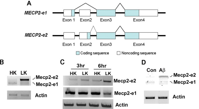
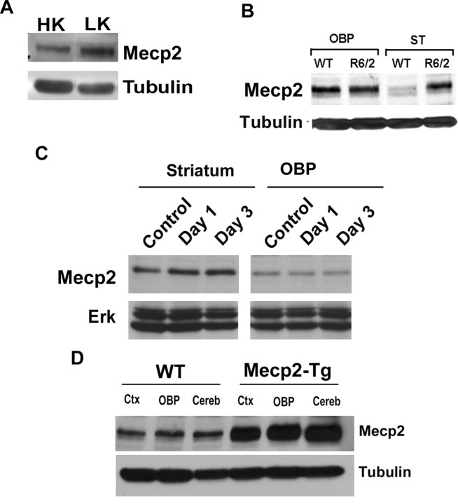
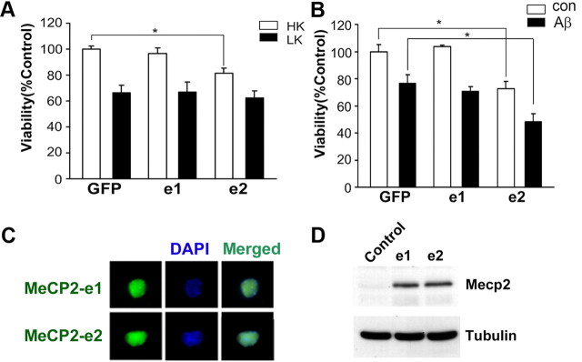
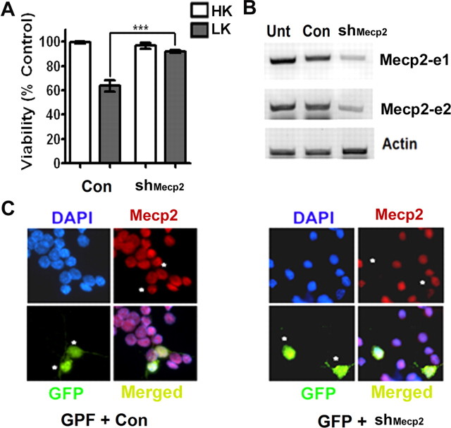
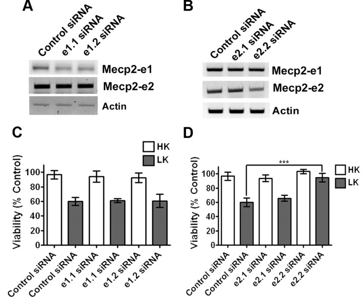
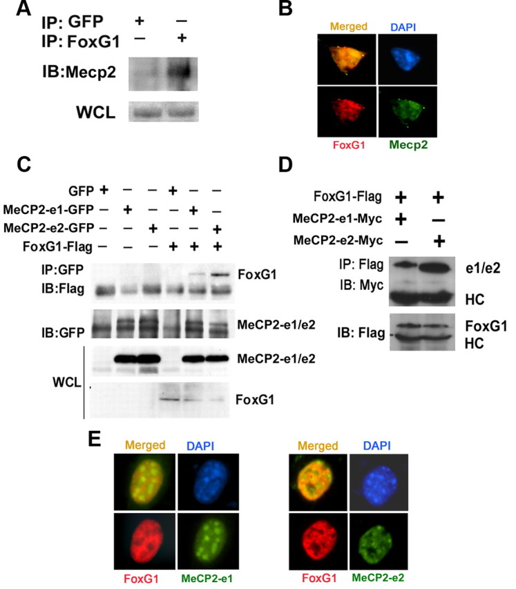
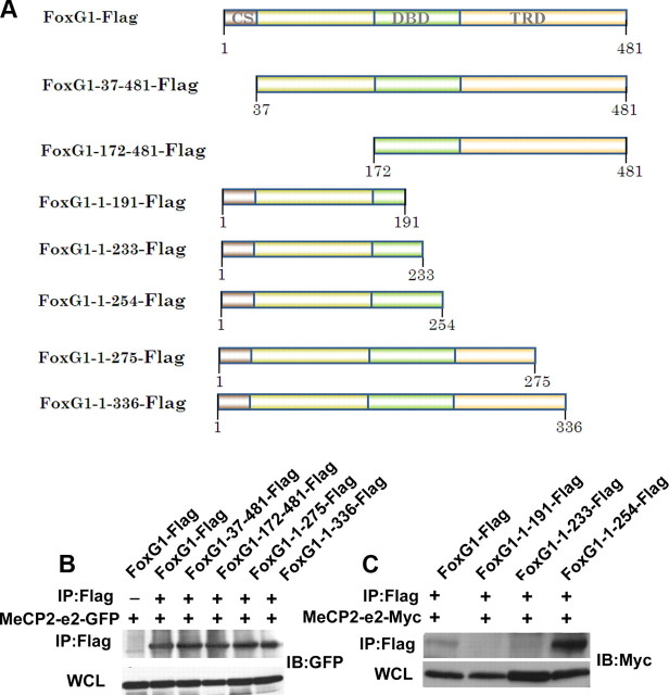
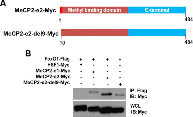
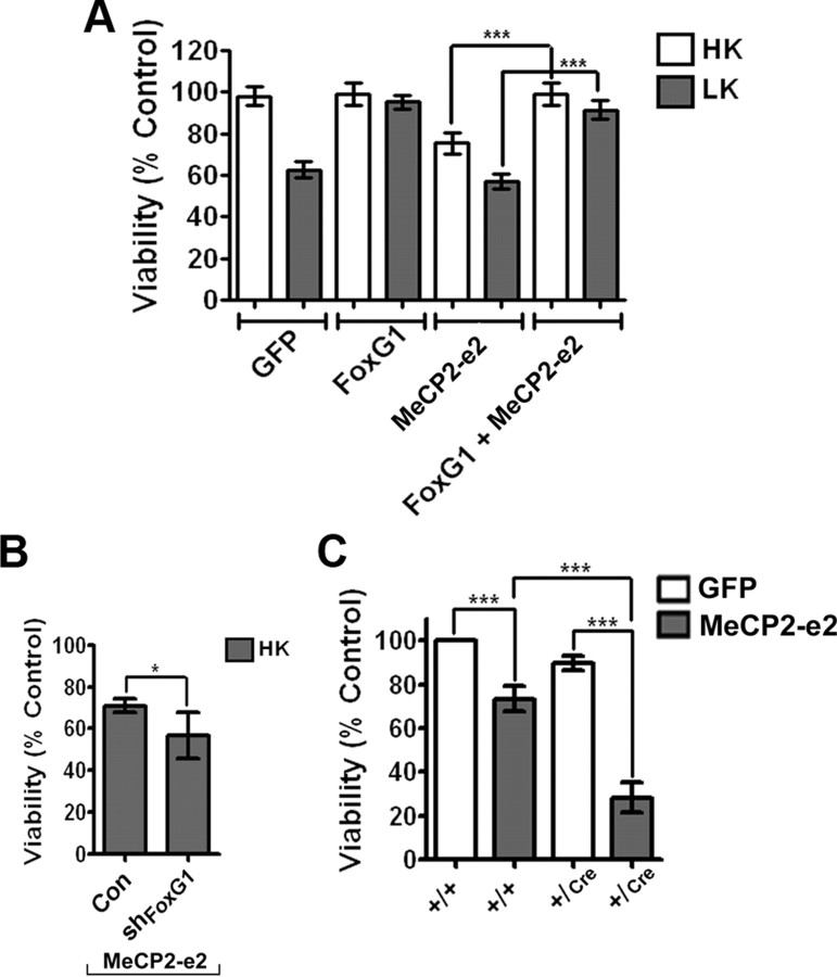
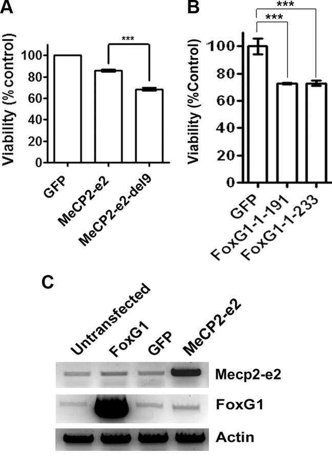
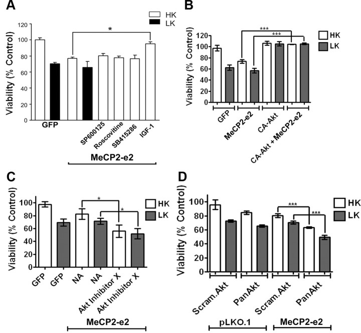
The methyl-CpG binding protein 2 (MeCP2) is a widely expressed protein, the mutations of which cause Rett syndrome. The level of MeCP2 is highest in the brain where it is expressed selectively in mature neurons. Its functions in postmitotic neurons are not known. The MeCP2 gene is alternatively spliced to generate two proteins with different N termini, designated as MeCP2-e1 and MeCP2-e2. The physiological significance of these two isoforms has not been elucidated, and it is generally assumed they are functionally equivalent. We report that in cultured cerebellar granule neurons induced to die by low potassium treatment and in Aβ-treated cortical neurons, Mecp2-e2 expression is upregulated whereas expression of the Mecp2-e1 isoform is downregulated. Knockdown of Mecp2-e2 protects neurons from death, whereas knockdown of the e1 isoform has no effect. Forced expression of MeCP2-e2, but not MeCP2-e1, promotes apoptosis in otherwise healthy neurons. We find that MeCP2-e2 interacts with the forkhead protein FoxG1, mutations of which also cause Rett syndrome. FoxG1 has been shown to promote neuronal survival and its downregulation leads to neuronal death. We find that elevated FoxG1 expression inhibits MeCP2-e2 neurotoxicity. MeCP2-e2 neurotoxicity is also inhibited by IGF-1, which prevents the neuronal death-associated downregulation of FoxG1 expression, and by Akt, the activation of which is necessary for FoxG1-mediated neuroprotection. Finally, MeCP2-e2 neurotoxicity is enhanced if FoxG1 expression is suppressed or in neurons cultured from FoxG1-haplodeficient mice. Our results indicate that Mecp2-e2 promotes neuronal death and that this activity is normally inhibited by FoxG1. Reduced FoxG1 expression frees MecP2-e2 to promote neuronal death.


Figures

Figure 1.
Figure 1.
Expression of Mecp2 isoforms in neurons primed to undergo apoptosis. A, Schematic representation of Mecp2–e1 and Mecp2–e2 isoforms. Mecp2–e1 isoform comprises exons 1, 3, and 4. Mecp2–e2 isoform comprises exons 2, 3, and 4. B, RT-PCR analysis of RNA prepared from cerebellar granule neurons (CGNs) treated with high potassium (HK) or low potassium (LK) for 4 h was performed with primers recognizing both e1 and e2 isoforms. β-actin served as the loading control. Mecp2–e2 isoform is upregulated in LK conditions. C, RT-PCR analysis of RNA isolated from 3 and 6 h HK- and LK-treated CGNs using primers specific for e1 or e2 isoform. Mecp2–e2 isoform is upregulated in LK conditions. D, RT-PCR analysis of RNA isolated from cortical neurons treated with 10 μm Aβ for 6 h. Primers recognizing both Mecp2 e1 and e2 isoforms were used. Mecp2–e2 isoform is upregulated in cortical neurons treated with Aβ.
Figure 2.
Figure 2.
Expression of Mecp2 isoforms in neurons primed to undergo apoptosis and in mouse models of neurodegeneration. A, Western blot analysis of Mecp2 protein in high potassium (HK)- or low potassium (LK)-treated cerebellar granule neurons (CGNs). The level of Mecp2 is upregulated in LK. B, Lysates were prepared from the striatum (ST) and all other brain parts (OBP) combined of 13-week-old R6/2 mice and nontransgenic littermates (WT), and subjected to Western blot analysis using an MeCP2 antibody. Tubulin served as the loading control. The level of Mecp2 is upregulated in the ST of R6/2 mice. C, Western blot analysis of ST and OBP of 10-week-old mice injected with either saline (control) or with 3-NP for 1 and 3 d. The blot was probed with an MeCP2 antibody. Total Erk served as the loading control. The level of Mecp2 was upregulated in the ST of 3-NP-injected mice as opposed to the saline-injected mice. D, Protein lysates obtained from the cortex (ctx), cerebellum (cereb), or the remainder of OBP of wild-type and Mecp2-overexpressing transgenic mice were subjected to Western blot analysis with an MeCP2 antibody. The elevated expression of the Mecp2 transgene is clearly detected by the antibody. In the brain part of the transgenic mice, Mecp2 was produced at least 3 times more than its wild-type littermate.
Figure 3.
Figure 3.
MeCP2–e2 isoform but not MeCP2–e1 isoform promotes neuronal death. A, Viability of CGNs transfected with GFP, MeCP2–e1-GFP (e1), or MeCP2–e2-GFP (e2) and switched to high potassium (HK) or low potassium (LK) medium for 24 h. The viability was normalized to GFP-transfected cultures treated with HK. MeCP2–e2 killed healthy neurons treated with HK. B, Viability of cortical neurons transfected with GFP, e1, or e2 and treated with no additives (con) or 10 μm Aβ for 48 h. Survival is normalized to GFP-transfected cortical neurons treated with no additives (con). MeCP2–e2 killed neurons treated with no additives (Con) as well as neurons treated with Aβ. C, CGNs transfected with MeCP2–e1 and MeCP2–e2 were subjected to immunocytochemistry using antibody against GFP. MeCP2–e1 and MeCP2–e2 were expressed at similar levels, as evident through immunocytochemistry. DAPI staining was performed to identify the nucleus of the cells. D, Western blot using MeCP2 antibody showing MeCP2–e1-GFP and MeCP2–e2-GFP isoform are expressed similarly in HEK293T cells. Tubulin served as the loading control. Control indicates untransfected HEK293T cells.
Figure 4.
Figure 4.
Effect of suppression of Mecp2–e1 and Mecp2–e2 expression in neurons using shRNA. A, Viability of CGNs transfected with control shRNA (Con) or shMecp2. Viability of transfected neurons was quantified and normalized to the viability of control cultures (Con-transfected cells). Knocking down endogenous levels of Mecp2 increased viability of neurons treated with LK. B, RT-PCR analysis of endogenous Mecp2 expression in HT22 cells that were either untransfected (Unt) or transfected with control shRNA (Con) or shMecp2. shMecp2 was able to knock down endogenous expression of both the isoforms of Mecp2. C, Immunocytochemistry using Mecp2 antibody showing knockdown of endogenous Mecp2 in neurons cotransfected with GFP and shMecp2 (indicated by asterisk). In neurons cotransfected with GFP and Con (indicated by asterisk) the level of endogenous Mecp2 is not reduced.
Figure 5.
Figure 5.
Effect of suppression of Mecp2–e1 and Mecp2–e2 expression in neurons using siRNA. A, RT-PCR analysis of endogenous Mecp2 expression in HT22 cells that were transfected with two different siRNAs (e1.1 and e1.2) against Mecp2–e1 isoform as indicated in the figure. The siRNA was able to knock down endogenous Mecp2–e1 isoform. B, RT-PCR analysis of endogenous Mecp2 expression in HT22 cells that were transfected with two differenet siRNAs (e2.1 and e2.1) against Mecp2–e2 isoform. One of the siRNAs, e2.2 siRNA, was able to knock down endogenous Mecp2–e2 isoform. C, Viability of CGNs cotransfected with GFP and siRNA (control or Mecp2–e1 specific). Viability was normalized to CGNs cotransfected with control siRNA and GFP. Knocking down endogenous Mecp2–e1 had no effect on neuronal survivability. D, Viability of CGNs cotransfected with GFP and siRNA (control or Mecp2–e2 specific). Viability was normalized to CGNs cotransfected with control siRNA and GFP. Knocking down endogenous Mecp2–e2 protected neurons against LK-mediated cell death.
Figure 6.
Figure 6.
Analysis of interaction between FoxG1 and Mecp2. A, CGN lysates were subjected to coimmunoprecipitation using FoxG1 antibody. The immunoprecipitate was analyzed by Western blot using MeCP2 antibody. FoxG1 was able to pull down Mecp2; however, GFP was unable to pull down Mecp2. An aliquot of the WCL (whole cell lysate) was also analyzed for Mecp2. B, Immunocytochemical analysis of CGNs using antibodies against FoxG1 and Mecp2. Mecp2 and FoxG1 colocalize. C, Lysates from HEK293T cells transfected with GFP, MeCP2–e1-GFP, MeCP2–e2-GFP, and Flag-FoxG1 plasmids as shown were immunoprecipitated using GFP antibody. Immunoblotting (IB) was performed using Flag antibody (top) and then reprobed with GFP antibody (bottom). Western blotting of WCL using GFP and Flag antibodies is also shown. FoxG1 interacts with both the isoforms of MeCP2 with a greater affinity for MeCP2–e2 isoform IP indicates immunoprecipitation. D, Lysates from HEK293T cells cotransfected with FoxG1-Flag and MeCP2–e1-Myc or MeCP2–e2-Myc were immunoprecipitated using Flag antibody. The immunoprecipitate was subjected to immunoblotting with Myc antibody (top). The blot was reprobed with Flag antibody (bottom). FoxG1 interacted with MeCP2–e2 isoform more strongly than the MeCP2–e1 isoform. E, HT22 cells were cotransfected with FoxG1-Flag and either MeCP2–e1-GFP or MeCP2–e2-GFP followed by immunocytochemistry using Flag and GFP antibodies. MeCP2–e2 and FoxG1 colocalize more than MeCP2–e1 and FoxG1.
Figure 7.
Figure 7.
Analysis of interaction between FoxG1 and MeCP2–e2. A, Schematic representation of the deletion constructs of FoxG1 used in this study. CS, Conserved sequence; DBD, DNA binding domain; TRD, transcriptional repression domain. B, Coimmunoprecipitation analysis of deletion constructs of FoxG1 (FoxG1-37–481, FoxG1-172–481, FoxG1-1–275, and FoxG1-1–336) and MeCP2–e2-GFP. The immunoprecipitation (IP) was performed with Flag antibody and immunoblotting (IB) was performed with GFP antibody. C, Coimmunoprecipitation analysis of deletion constructs of FoxG1 (FoxG1-1–191, FoxG1-1–233,and FoxG1-1–254) and MeCP2–e2-Myc. The immunoprecipitation was performed with Flag antibody and immunoblotting was performed with Myc antibody. FoxG1-1–191 and FoxG1-1–233 do not interact with MeCP2–e2.
Figure 8.
Figure 8.
Analysis of interaction between FoxG1 and MeCP2–e2. A, Schematic representation of deletion construct of MeCP2–e2. B, Coimmunoprecipitation analysis of MeCP2–e2-del9-Myc with FoxG1. Immunoprecipitation (IP) was performed with Flag antibody and immunoblotting (IB) was performed with Myc antibody. MeCP2–e2-del9-Myc does not interact with FoxG1-Flag like MeCP2–e2.
Figure 9.
Figure 9.
MeCP2–e2 neurotoxicity is inhibited by FoxG1. A, Viability of CGNs transfected with GFP, FoxG1-Flag, and MeCP2–e2-GFP either by themselves or in combination and treated with HK or LK for 24 h. Viability was normalized to neurons transfected with GFP and treated with HK. FoxG1 can rescue against MeCP2–e2-mediated neuronal death. B, CGNs were cotransfected with MeCP2–e2 and either pLKO.1 (Con) or shFoxG1. The effectiveness of shFoxG1 (also referred to as #1746 in Dastidar et al., 2011) to suppress FoxG1 expression has previously been demonstrated (Dastidar et al., 2011). Viability of transfected cells identified by GFP immunoreactivity was quantified and compared with control shRNA and MeCP2–e2-GFP transfected neurons treated with HK. C, Viability of CGNs cultured from FoxG1+/+ and FoxG1+/Cre mice transfected with GFP or MeCP2–e2- GFP and treated with HK for 14 h. Viability was normalized to FoxG1+/+ CGNs transfected with GFP. Neurotoxicity is higher in neurons harvested from FoxG1+/Cre mice as compared with FoxG1+/+ neurons.
Figure 10.
Figure 10.
MeCP2–e2 neurotoxicity is inhibited by FoxG1. A, Viability of neurons transfected with GFP, MeCP2–e2-Myc, and MeCP2–e2-del9-Myc and treated with HK for 24 h. Viability of neurons was normalized to CGNs transfected with GFP and treated with HK. MeCP2–e2-del9 killed healthy neurons more than MeCP2–e2. B, Viability of neurons transfected with GFP, FoxG1-1–191-Flag, and FoxG1-1–233- Flag and treated with HK for 24 h. Viability of neurons was normalized to CGNs transfected with GFP and treated with HK. Both the deleted constructs of FoxG1 killed neurons. C, RT-PCR analysis of HT22 cells transfected with FoxG1, GFP, and MeCP2–e2. Overexpression of FoxG1 did not affect endogenous mRNA levels of Mecp2, and similarly overexpression of MeCP2–e2 did not affect the endogenous mRNA levels of FoxG1.
Figure 11.
Figure 11.
IGF-1 blocks MeCP2–e2-mediated neurotoxicity. A, Viability of CGNs transfected with GFP or MeCP2–e2- GFP and switched to HK or LK medium or LK medium supplemented with inhibitors (SP600125, roscovitine, or SB415286) or with IGF-1. Viability of transfected cells was quantified by DAPI staining and normalized to GFP-transfected cultures treated with HK (control). IGF-1 protected against MeCP2–e2-mediated neuronal death. B, Viability of CGNs transfected with GFP, MeCP2–e2-GFP, and CA-Akt-HA either by themselves or in combination and treated with HK or LK for 24 h. Viability was normalized to neurons transfected with GFP and treated with HK. CA-Akt can rescue against MeCP2–e2-mediated neuronal death. C, Viability of CGNs transfected with GFP or MeCP2–e2 and treated with HK or LK or HK or LK with Akt Inhibitor X. Viability of transfected neurons was normalized to GFP transfected neurons treated with HK. Blocking Akt increased MeCP2–e2 toxicity. D, Viability of CGNs cotransfected with pLKO.1 and either Scrambled Akt (Scram.Akt) or Pan Akt shRNA and MeCP2–e2 with either Scrambled Akt or Pan Akt shRNA. Viability was normalized to CGNs transfected with pLKO.1 and Scrambled Akt. Bringing down the levels of endogenous Akt increased MeCP2–e2-mediated neuronal death. NA, no additive.

Comment in

References

    1. Ariani F, Hayek G, Rondinella D, Artuso R, Mencarelli MA, Spanhol-Rosseto A, Pollazzon M, Buoni S, Spiga O, Ricciardi S, Meloni I, Longo I, Mari F, Broccoli V, Zappella M, Renieri A. FOXG1 is responsible for the congenital variant of Rett syndrome. Am J Hum Genet. 2008;83:89–93. - PMC - PubMed
    1. Bahi-Buisson N, Nectoux J, Girard B, Van Esch H, De Ravel T, Boddaert N, Plouin P, Rio M, Fichou Y, Chelly J, Bienvenu T. Revisiting the phenotype associated with FOXG1 mutations: two novel cases of congenital Rett variant. Neurogenetics. 2010;11:241–249. - PubMed
    1. Bardai FH, D'Mello SR. Selective toxicity by HDAC3 in neurons: regulation by akt and GSK3β. J Neurosci. 2011;31:1746–1751. - PMC - PubMed
    1. Beal MF, Brouillet E, Jenkins BG, Ferrante RJ, Kowall NW, Miller JM, Storey E, Srivastava R, Rosen BR, Hyman BT. Neurochemical and histological characterization of striatal excitotoxic lesions produced by the mitochondrial toxin 3-Nitropropionic acid. J Neurosci. 1993;13:4181–4192. - PMC - PubMed
    1. Björkblom B, Vainio JC, Hongisto V, Herdegen T, Courtney MJ, Coffey ET. All JNKs can kill, but nuclear localization is critical for neuronal death. J Biol Chem. 2008;283:19704–19713. - PubMed

Publication types

MeSH terms

LinkOut - more resources