

Ultrastructure of synapses in the mammalian brain
- PMID: 22357909
- PMCID: PMC3331701
- DOI: 10.1101/cshperspect.a005587
Ultrastructure of synapses in the mammalian brain
Abstract
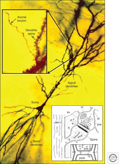
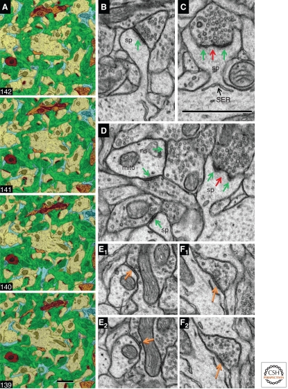
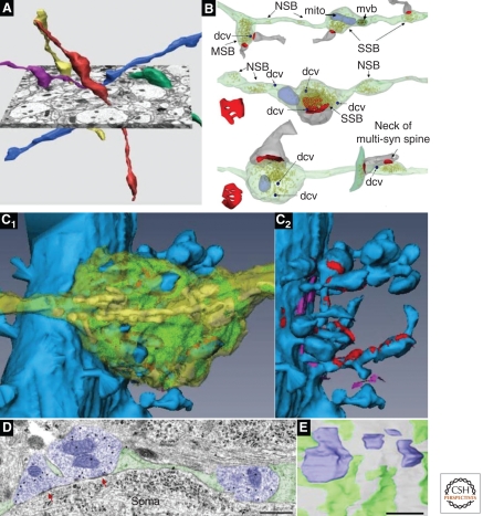
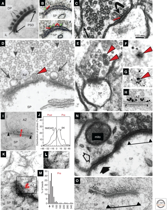
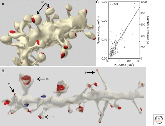
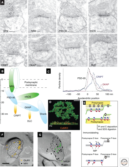
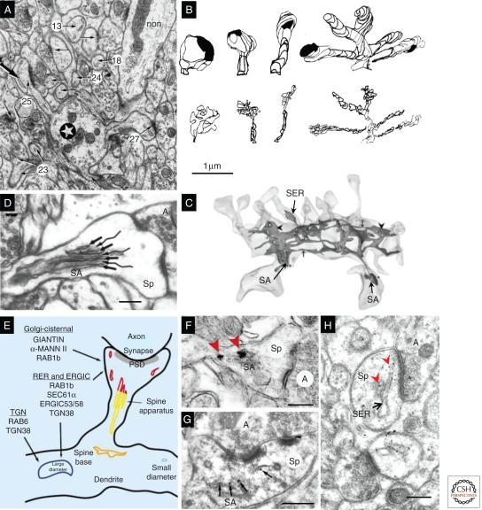
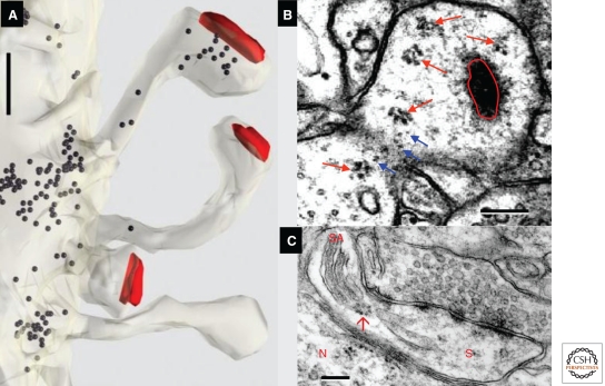
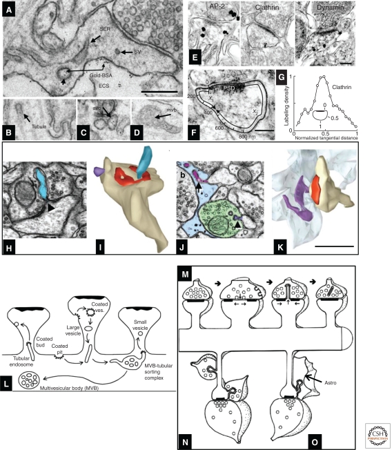
The morphology and molecular composition of synapses provide the structural basis for synaptic function. This article reviews the electron microscopy of excitatory synapses on dendritic spines, using data from rodent hippocampus, cerebral cortex, and cerebellar cortex. Excitatory synapses have a prominent postsynaptic density, in contrast with inhibitory synapses, which have less dense presynaptic or postsynaptic specializations and are usually found on the cell body or proximal dendritic shaft. Immunogold labeling shows that the presynaptic active zone provides a scaffold for key molecules involved in the release of neurotransmitter, whereas the postsynaptic density contains ligand-gated ionic channels, other receptors, and a complex network of signaling molecules. Delineating the structure and molecular organization of these axospinous synapses represents a crucial step toward understanding the mechanisms that underlie synaptic transmission and the dynamic modulation of neurotransmission associated with short- and long-term synaptic plasticity.
Figures
References
-
- Ahmari SE, Smith SJ 2002. Knowing a nascent synapse when you see it. Neuron 34: 333–336 - PubMed
-
- Amaral DG, Dent JA 1981. Development of the mossy fibers of the dentate gyrus: I. A light and electron microscopic study of the mossy fibers and their expansions. J Comp Neurol 195: 51–86 - PubMed
-
- Baude A, Bleasdale C, Dalezios Y, Somogyi P, Klausberger T 2007. Immunoreactivity for the GABAA receptor α1 subunit, somatostatin and Connexin36 distinguishes axoaxonic, basket, and bistratified interneurons of the rat hippocampus. Cereb Cortex 17: 2094–2107 - PubMed
Publication types
MeSH terms
LinkOut - more resources
Full Text Sources