

Rapid pediatric cardiac assessment of flow and ventricular volume with compressed sensing parallel imaging volumetric cine phase-contrast MRI
- PMID: 22358022
- PMCID: PMC3515670
- DOI: 10.2214/AJR.11.6969
Rapid pediatric cardiac assessment of flow and ventricular volume with compressed sensing parallel imaging volumetric cine phase-contrast MRI
Abstract
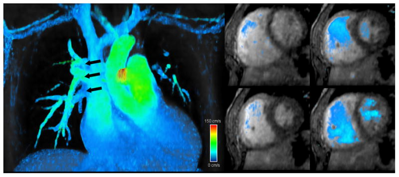
Objective: The quantification of cardiac flow and ventricular volumes is an essential goal of many congenital heart MRI examinations, often requiring acquisition of multiple 2D phase-contrast and bright-blood cine steady-state free precession (SSFP) planes. Scan acquisition, however, is lengthy and highly reliant on an imager who is well-versed in structural heart disease. Although it can also be lengthy, 3D time-resolved (4D) phase-contrast MRI yields global flow patterns and is simpler to perform. We therefore sought to accelerate 4D phase contrast and to determine whether equivalent flow and volume measurements could be extracted.
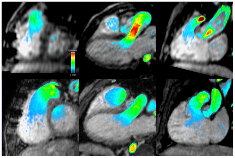
Materials and methods: Four-dimensional phase contrast was modified for higher acceleration with compressed sensing. Custom software was developed to process 4D phase-contrast images. We studied 29 patients referred for congenital cardiac MRI who underwent a routine clinical protocol, including cine short-axis stack SSFP and 2D phase contrast, followed by contrast-enhanced 4D phase contrast. To compare quantitative measurements, Bland-Altman analysis, paired Student t tests, and F tests were used.
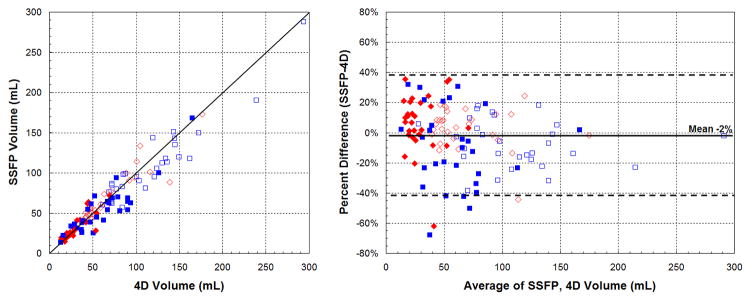
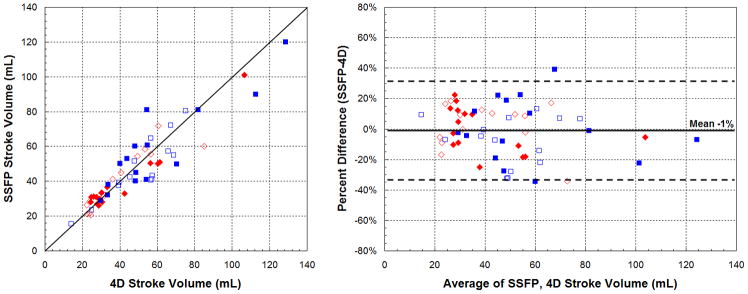
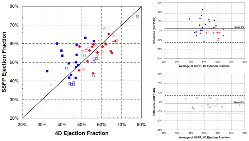
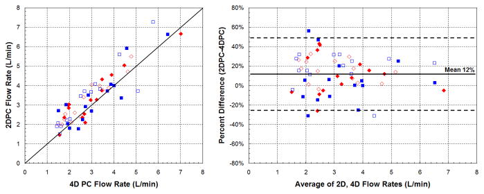
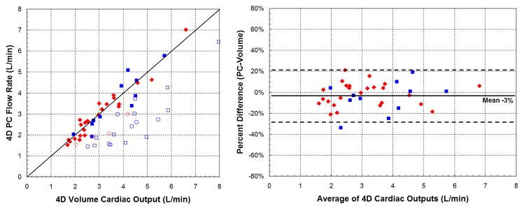
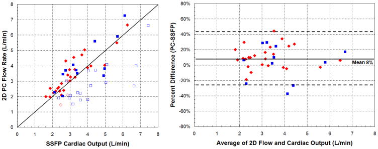
Results: Ventricular end-diastolic, end-systolic, and stroke volumes obtained from 4D phase contrast and SSFP were well correlated (ρ = 0.91-0.95; r(2) = 0.83-0.90), with no statistically significant difference. Ejection fractions were well correlated in a subpopulation that underwent higher-resolution compressed-sensing 4D phase contrast (ρ = 0.88; r(2) = 0.77). Four-dimensional phase contrast and 2D phase contrast flow rates were also well correlated (ρ = 0.90; r(2) = 0.82). Excluding ventricles with valvular insufficiency, cardiac outputs derived from outlet valve flow and stroke volumes were more consistent by 4D phase contrast than by 2D phase contrast and SSFP.
Conclusion: Combined parallel imaging and compressed sensing can be applied to 4D phase contrast. With custom software, flow and ventricular volumes may be extracted with comparable accuracy to SSFP and 2D phase contrast. Furthermore, cardiac outputs were more consistent by 4D phase contrast.
Figures
References
-
- Sechtem U, Pflugfelder PW, Gould RG, Cassidy MM, Higgins CB. Measurement of right and left ventricular volumes in healthy individuals with cine MR imaging. Radiology. 1987;163:697–702. - PubMed
-
- Longmore DB, Klipstein RH, Underwood SR, et al. Dimensional accuracy of magnetic resonance in studies of the heart. Lancet. 1985;1:1360–1362. - PubMed
-
- Carr JC, Simonetti O, Bundy J, Li D, Pereles S, Finn JP. Cine MR Angiography of the Heart with Segmented True Fast Imaging with Steady-State Precession1. Radiology. 2001;219:828–834. - PubMed
-
- Devos D, Kilner P. Calculations of cardiovascular shunts and regurgitation using magnetic resonance ventricular volume and aortic and pulmonary flow measurements. European Radiology. 2010;20:410–421. - PubMed
-
- Higgins CB, Sakuma H. Heart disease: functional evaluation with MR imaging. Radiology. 1996;199:307–315. - PubMed
Publication types
MeSH terms
Substances
Grants and funding
LinkOut - more resources
Full Text Sources
Other Literature Sources
Medical
