Multicenter Study
. 2012 Jun;81(11):1140-8.
doi: 10.1038/ki.2012.7. Epub 2012 Feb 22.

Urine risk factors in children with calcium kidney stones and their siblings

Affiliations
Multicenter Study

Urine risk factors in children with calcium kidney stones and their siblings

Kristin J Bergsland et al. Kidney Int. 2012 Jun.

Abstract

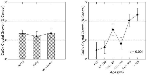
Calcium nephrolithiasis in children is increasing in prevalence and tends to be recurrent. Although children have a lower incidence of nephrolithiasis than adults, its etiology in children is less well understood; hence, treatments targeted for adults may not be optimal in children. To better understand metabolic abnormalities in stone-forming children, we compared chemical measurements and the crystallization properties of 24-h urine collections from 129 stone formers matched to 105 non-stone-forming siblings and 183 normal, healthy children with no family history of stones, all aged 6 to 17 years. The principal risk factor for calcium stone formation was hypercalciuria. Stone formers have strikingly higher calcium excretion along with high supersaturation for calcium oxalate and calcium phosphate, and a reduced distance between the upper limit of metastability and supersaturation for calcium phosphate, indicating increased risk of calcium phosphate crystallization. Other differences in urine chemistry that exist between adult stone formers and normal individuals such as hyperoxaluria, hypocitraturia, abnormal urine pH, and low urine volume were not found in these children. Hence, hypercalciuria and a reduction in the gap between calcium phosphate upper limit of metastability and supersaturation are crucial determinants of stone risk. This highlights the importance of managing hypercalciuria in children with calcium stones.


Figures

Figure 1
Figure 1. Urine stone risk factors
Mean 24-hr excretions of calcium (top left), oxalate (top middle), phosphorus (top right), and citrate (bottom right), as well as urine volume (bottom left) and pH (bottom middle), are shown for children with calcium stones, their non stone forming siblings, and normal healthy children. Values are means ± SEM, adjusted for gender, age and body surface area. Significant differences between subject types were observed only for calcium excretion: *p < 0.0001, stone former vs. sibling and stone former vs. normal; #p < 0.03, sibling vs. normal.
Figure 2
Figure 2. Calcium excretion and stone formation
Daily calcium excretion is expressed per 1.73 m2 body surface area, adjusted for gender and age. A quantile plot showing the distribution of data for all subjects is at left, and means ± SEM by subject group at right. Stone formers are divided into two groups: those who have had 1–2 stones (79 subjects; 34 boys, 45 girls) and those with >2 stones (50 subjects; 22 boys, 28 girls). Significant differences are as follows: *p < 0.0001, >2 stones vs. sibling, >2 stones vs. normal and 1–2 stones vs. normal; p < 0.01, >2 stones vs. 1–2 stones; #p < 0.03, 1–2 stones vs. sibling and sibling vs. normal. Stone formers and their siblings excreted more calcium than their normal counterparts, and calcium excretion was higher in patients with more stones (right panel, p<0.0001 for the regression).
Figure 3
Figure 3. Urine calcium, oxalate and citrate by gender and age
Daily excretions of calcium (left panels), oxalate (middle panels) and citrate (right panels) are expressed per 1.73 m2 body surface area in boys (top panels) and girls (bottom panels) by age sextile. Values are means ± SEM. Oxalate declines significantly with age in girls (lower middle panel) and citrate declines with age in boys (upper right panel), p<0.001 for both regressions.
Figure 4
Figure 4. Urine SS and ULM for CaOx and CaP
CaOx SS and CaP SS by subject group are shown as black circles at lower ends of each bar in the left and right panels, respectively; the upper limit of metastability (ULM), the supersaturation required to experimentally induce crystallization, are the grey circles at upper ends of each bar. Values are means ± SEM. In the lower panels, stone formers are divided into two groups: those who have had 1–2 stones and those with >2 stones (see Figure 2). Hatched bars represent the distance between ambient SS and ULM, an indicator of the capacity of the urine to resist crystallization. CaOx ULM rose with CaOx SS across the groups from normal to stone former (upper left panel) and from normal through sibling to progressively more active stone former (lower left panel) so that the ULM – SS distance (length of cross hatched bars) remained constant. In stone formers, the CaP ULM – SS distance fell dramatically (upper right panel, length of cross hatched bars), most notably among those with >2 stones (lower right panel), suggesting that the CaP ULM – SS distance may be an important determinant of stone risk. Significant differences in SS or ULM: * p < 0.001 vs. NN; # p<0.01 vs. NS; + p<0.05 vs. NN; § p<0.05 vs. NN.
Figure 5
Figure 5. Calcium oxalate crystal growth inhibition
Crystal growth is shown as mean ± SEM percent control growth for children grouped by subject type (left), or by age sextile (right). Results are adjusted for gender and body surface area. Higher values are closer to the control containing no inhibitory urine protein, and indicate weaker inhibition. Crystal growth inhibition is not different by subject type but diminishes with age (p<0.001 for the regression).

Comment in

Similar articles

Cited by

References

    1. Worcester EM, Coe FL. Clinical practice. Calcium kidney stones. N Engl J Med. 2010;363:954–963. - PMC - PubMed
    1. Pak CY. Medical management of urinary stone disease. Nephron Clin Pract. 2004;98:c49–c53. - PubMed
    1. Coe FL, Parks JH, Moore ES. Familial idiopathic hypercalciuria. N Engl J Med. 1979;300:337–340. - PubMed
    1. Evan AP. Physiopathology and etiology of stone formation in the kidney and the urinary tract. Pediatr Nephrol. 2010;25:831–841. - PMC - PubMed
    1. Coe FL, Parks JH. New insights into the pathophysiology and treatment of nephrolithiasis: new research venues. J Bone Miner Res. 1997;12:522–533. - PubMed

Publication types

MeSH terms

LinkOut - more resources