. 2012 Feb 22;482(7386):501-6.
doi: 10.1038/nature10829.

Structural basis of highly conserved ribosome recycling in eukaryotes and archaea

Affiliations

Structural basis of highly conserved ribosome recycling in eukaryotes and archaea

Thomas Becker et al. Nature. .

Abstract

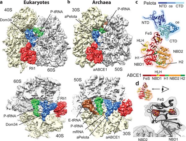
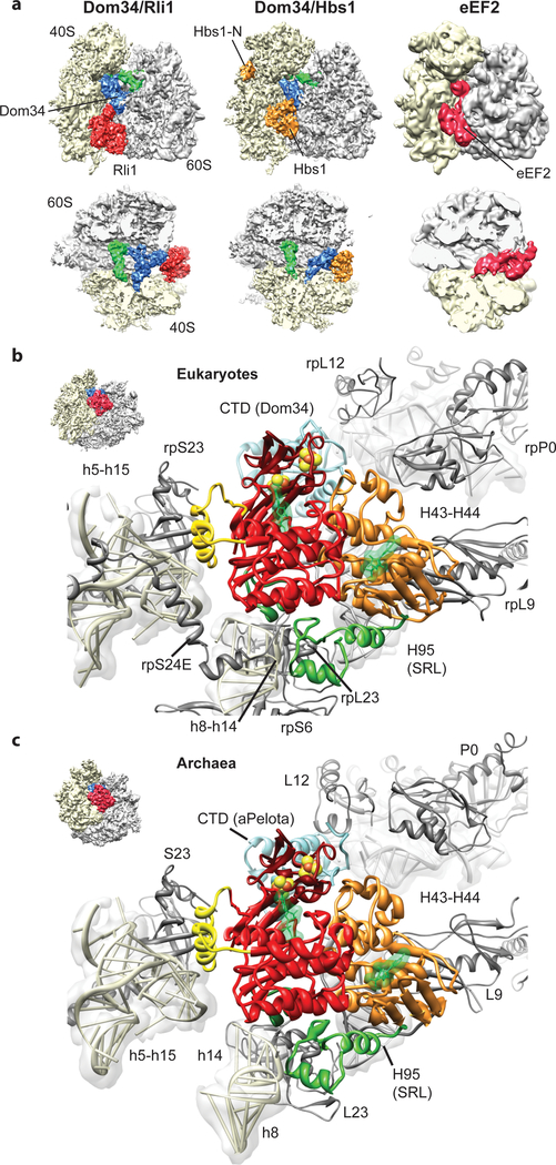
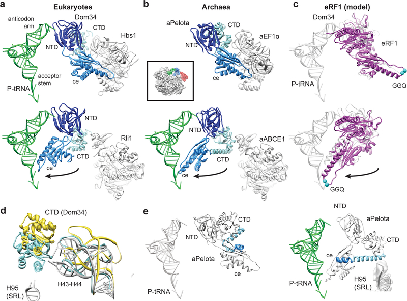
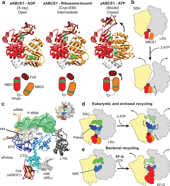
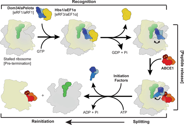
Ribosome-driven protein biosynthesis is comprised of four phases: initiation, elongation, termination and recycling. In bacteria, ribosome recycling requires ribosome recycling factor and elongation factor G, and several structures of bacterial recycling complexes have been determined. In the eukaryotic and archaeal kingdoms, however, recycling involves the ABC-type ATPase ABCE1 and little is known about its structural basis. Here we present cryo-electron microscopy reconstructions of eukaryotic and archaeal ribosome recycling complexes containing ABCE1 and the termination factor paralogue Pelota. These structures reveal the overall binding mode of ABCE1 to be similar to canonical translation factors. Moreover, the iron-sulphur cluster domain of ABCE1 interacts with and stabilizes Pelota in a conformation that reaches towards the peptidyl transferase centre, thus explaining how ABCE1 may stimulate peptide-release activity of canonical termination factors. Using the mechanochemical properties of ABCE1, a conserved mechanism in archaea and eukaryotes is suggested that couples translation termination to recycling, and eventually to re-initiation.


Figures

Fig. 1:
Fig. 1:. The ribosome-bound Pelota-ABCE1 complex
(a, b) Cryo-EM reconstructions of the eukaryotic SL-RNC-Dom34-Rli1 and the archaeal 70S-aPelota-aABCE1 complex at 7.2 Å and 6.6 Å resolution, respectively. Extra densities were observed for Dom34/aPelota and Rli1/aABCE1 in the canonical factor binding site as well as for P-site tRNA, E-site tRNA and mRNA. The upper section represents side views, the lower section top views, where large and small subunits were cut. (c) Homology model for ribosome-bound Pelota and ABCE1 in transparent density. The individual domains are color-coded as in the schematic representation of domain organization. N-terminal Domain (NTD), central domain (ce) and C-terminal domain (CTD) are indicated; FeS indicates iron-sulfur cluster domain; NBD1 and 2 indicate nucleotide binding domain 1 and 2; HLH indicates helix-loop-helix motif; H1 and H2 indicates hinge 1 and hinge 2 domain. (d) Zoom on the FeS domain of aABCE1. The density for the two [4Fe-4S]2+ is displayed in red mesh at high contour level.
Fig. 2:
Fig. 2:. Interaction of Pelota and ABCE1 with the ribosome.
(a) Comparison of the SL-RNC-Dom34-Rli1 cryo-EM map with the SL-RNC-Dom34-Hbs1 and the 80S-eEF2 maps. Views are as in Fig. 1a and 1b. (b, c) Interactions of ABCE1 with the eukaryotic (b) and the archaeal (c) ribosome. The view is indicated by a thumbnail. The domain color code is as in Fig. 1c.
Fig. 3:
Fig. 3:. Domain movements in Pelota and eRF1
(a) Comparison of the ribosome-bound Dom34 conformation in complex with Hbs1 (top section) and Rli1 (lower section). (b) Comparison of the aPelota-aEF1α crystal structure with the ribosome-bound aPelota in complex with aABCE1. The central domain of Pelota swings out towards the P-site tRNA. (c) Models for eRF1 before and after the suggested movement of the central domain. (d) Conformation of the Dom34 CTD and the stalk base rRNA (H43-H44) when bound to Hbs1 (yellow) and to Rli1 (blue). rRNA conformation without factors bound is shown in grey. (e) In aPelota three separate small helices refold into a long α-helix during movement of the central domain bridging the CTD and the central domain.
Fig. 4:
Fig. 4:. Mechanochemical activity of ABCE1 on the ribosome
(a) Crystal structure of the open (ADP-bound) aABCE1, the cryo-EM structure of the ribosome-bound aABCE1 and homology model of the closed (ATP-bound) aABCE1 including schematic drawings. An asterisk indicates a contact between NBD2 and the FeS domain of aABCE1. (b) Ribosomal subunits may be dissociated by following the trajectory of aABCE1 domain closure upon ATP-binding. (c) Interactions of the aPelota NTD and central domain within the ribosome. (d) ABCE1 domain closure could lead to an allosteric cascade with the FeS domain acting as a bolt on the CTD of Pelota to rearrange the NTD and central domain of Pelota. This mechanism would be analogous to the splitting reaction in bacteria by RRF and EF-G as depicted in (e).
Fig. 5:
Fig. 5:. Scheme of archaeal and eukaryotic ribosome recycling bridging termination with initiation.
A translational GTPase (Hbs1/aEF1α/eRF3) delivers the factor, which recognizes stalled ribosomes (Pelota) or pre-termination complexes (eRF1/aRF1). After GTP hydrolysis, the GTPase dissociates and ABCE1 can bind. ABCE1 induces or stabilizes the swung-out conformation of Pelota (or RF1), which would lead to peptide release in case of termination. Ribosome splitting is induced after ATP-binding to ABCE1 and hydrolysis. In eukaryotes initiation factors can bind during the splitting reaction coupling ribosome recycling with re-initiation. After splitting ABCE1 stays associated with the small ribosomal subunit.

Similar articles

Cited by

References

    1. Winzeler EA et al. Functional characterization of the S. cerevisiae genome by gene deletion and parallel analysis. Science 285, 901–906 (1999). - PubMed
    1. Coelho CM et al. Growth and cell survival are unevenly impaired in pixie mutant wing discs. Development 132, 5411–5424 (2005). - PubMed
    1. Estevez AM, Haile S, Steinbuchel M, Quijada L & Clayton C. Effects of depletion and overexpression of the Trypanosoma brucei ribonuclease L inhibitor homologue. Mol. Biochem. Parasitol 133, 137–141 (2004). - PubMed
    1. Pisarev AV et al. The role of ABCE1 in eukaryotic posttermination ribosomal recycling. Mol. Cell 37, 196–210 (2010). - PMC - PubMed
    1. Pisareva VP, Skabkin MA, Hellen CU, Pestova TV & Pisarev AV Dissociation by Pelota, Hbs1 and ABCE1 of mammalian vacant 80S ribosomes and stalled elongation complexes. Embo J. (2011). - PMC - PubMed

Publication types

MeSH terms