. 2012 Feb 22;482(7386):552-6.
doi: 10.1038/nature10867.

Structure and dynamics of the M3 muscarinic acetylcholine receptor

Affiliations

Structure and dynamics of the M3 muscarinic acetylcholine receptor

Andrew C Kruse et al. Nature. .

Abstract

Acetylcholine, the first neurotransmitter to be identified, exerts many of its physiological actions via activation of a family of G-protein-coupled receptors (GPCRs) known as muscarinic acetylcholine receptors (mAChRs). Although the five mAChR subtypes (M1-M5) share a high degree of sequence homology, they show pronounced differences in G-protein coupling preference and the physiological responses they mediate. Unfortunately, despite decades of effort, no therapeutic agents endowed with clear mAChR subtype selectivity have been developed to exploit these differences. We describe here the structure of the G(q/11)-coupled M3 mAChR ('M3 receptor', from rat) bound to the bronchodilator drug tiotropium and identify the binding mode for this clinically important drug. This structure, together with that of the G(i/o)-coupled M2 receptor, offers possibilities for the design of mAChR subtype-selective ligands. Importantly, the M3 receptor structure allows a structural comparison between two members of a mammalian GPCR subfamily displaying different G-protein coupling selectivities. Furthermore, molecular dynamics simulations suggest that tiotropium binds transiently to an allosteric site en route to the binding pocket of both receptors. These simulations offer a structural view of an allosteric binding mode for an orthosteric GPCR ligand and provide additional opportunities for the design of ligands with different affinities or binding kinetics for different mAChR subtypes. Our findings not only offer insights into the structure and function of one of the most important GPCR families, but may also facilitate the design of improved therapeutics targeting these critical receptors.


Conflict of interest statement

The authors declare no competing financial interests

Figures

Figure 1
Figure 1. Major structural features of the M3 receptor
a, Analysis of muscarinic receptor sequences divides them into two classes. b, The overall structure of the M3 receptor (green) is similar to that of the M2 receptor (orange). The M3-bound ligand, tiotropium, is shown in spheres c, Comparison of the intracellular surfaces shows divergence in the cytoplasmic end of transmembrane helix 5. d, Comparison of the extracellular surfaces shows less deviation, with near perfect conservation of backbone fold of extracellular loops e, A solvent accessible surface for the M3 receptor bound to tiotropium (spheres) shows the receptor covering the ligand with a tyrosine lid (outlined in red). f, M3 receptor structure colored by sequence conservation among the five mAChR subtypes. Poorly conserved regions are shown with larger backbone diameter. The orthosteric and allosteric sites are indicated in blue and red, respectively, and the ligand tiotropium is shown in spheres.
Figure 2
Figure 2. Orthosteric binding sites of the M2 and M3 receptors
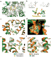
In all panels, the M3 receptor is green with its ligand tiotropium in yellow, while the M2 receptor and its ligand QNB are shown in orange and cyan, respectively. a, Tiotropium binding site in the M3 receptor. A 2Fo-Fc map contoured at 2Fo-Fc is shown in wire. b, Chemical structures of ligands. A red arrow indicates the tropane C3 atom used as a tracking landmark in Fig. 3. Superimposing the receptor structures reveals that the two ligands adopt highly similar poses (bottom). c, There is a Phe (M2)/Leu (M3) sequence difference between the M2 and M3 receptors near the binding site. d, This produces an enlarged binding pocket in the M3 receptor. e, A displacement of M3 Y5297.39 is seen. f, This may arise from a sequence difference at position 2.61 (Tyr80 in M2 and Phe124 in M3).
Figure 3
Figure 3. Molecular dynamics of ligand binding
Simulations suggest that the tiotropium binding/dissociation pathway for both receptors involves a metastable state in the extracellular vestibule. a, When tiotropium is pushed out of the binding pocket of M3, it pauses in the extracellular vestibule. Spheres represent positions of the ligand’s C3 tropane atom at successive points in time. b, When tiotropium is placed in solvent, it binds to the same site in the extracellular vestibule. Our simulations are insufficiently long for it to proceed into the orthosteric binding pocket; the agonist ACh, a much smaller molecule, bound spontaneously to the orthosteric site in similar simulations (see Supplementary Methods) c, Schematic free energy landscape for binding/dissociation. d, Common binding poses for tiotropium in the extracellular vestibule of M2 (orange) and of M3 (green). Non-conserved residues that contact the ligand are shown in thin sticks. The location of the orthosteric site is indicated by tiotropium in spheres.
Figure 4
Figure 4. G protein coupling specificity determinants
a, The M3 receptor shows displacement of TM5 relative to its position in M2, and a conserved tyrosine (M3 Tyr2505.58) adopts different positions in the two receptors. Four TM6 residues near TM5 (AALS in M3, VTIL in M2) have been shown to be important coupling specificity determinants. b, ICL2 is also divergent between the two structures. Four residues previously implicated as specificity determinants are shown, with residue numbers for M2 followed by M3 c, Plot of interhelical distances for crystallographically unique inactive GPCR structures published to date. Distances were measured between Cα atoms of TM5 residue 5.62 and TM3 residue 3.54 (x-axis), and TM5 residue 5.62 and TM6 residue 6.37 (y-axis). GPCRs cluster by coupling specificity, although squid rhodopsin is an exception. GPCRs coupling preferentially to Gi/o and those coupling to the homologous G protein Gt cluster together. d, Structural alignment of mammalian Gi/o-coupled and Gq/11-coupled receptor structures.

Comment in

References

    1. Loewi O. Uber humorale ubertragbarkeit der Herznervenwirkung. Pflugers Arch. 1921;189:239–242.
    1. Hulme EC, Birdsall NJM, Buckley NJ. Muscarinic receptor subtypes. Ann Rev Pharmacol and Toxicol. 1990;30:633–673. - PubMed
    1. Wess J. Molecular biology of muscarinic acetylcholine receptors. Crit Rev Neurobiol. 1996;10:69–99. - PubMed
    1. Caulfield MP, Birdsall NJM. International union of pharmacology. XVII Classification of muscarinic acetylcholine receptors. Pharmacol Rev. 1998;50 :279–290. - PubMed
    1. Conn PJ, Jones CK, Lindsley CW. Subtype-selective allosteric modulators of muscarinic receptors for the treatment of CNS disorders. Trends Pharmacol Sci. 2009;30:148–155. - PMC - PubMed

Publication types

Associated data