. 2012;8(2):e1002368.
doi: 10.1371/journal.pcbi.1002368. Epub 2012 Feb 16.

Dynamic energy landscapes of riboswitches help interpret conformational rearrangements and function

Affiliations

Dynamic energy landscapes of riboswitches help interpret conformational rearrangements and function

Giulio Quarta et al. PLoS Comput Biol. 2012.

Abstract

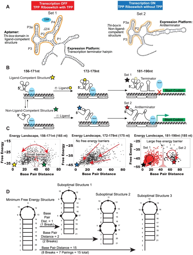
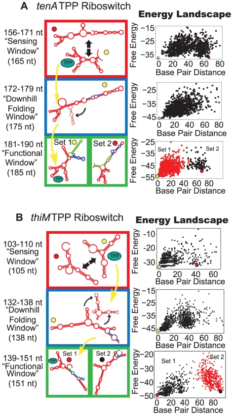
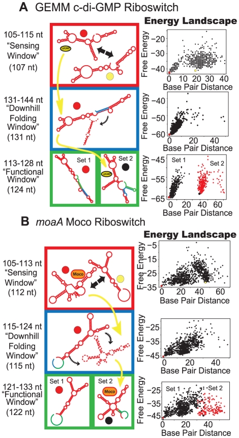
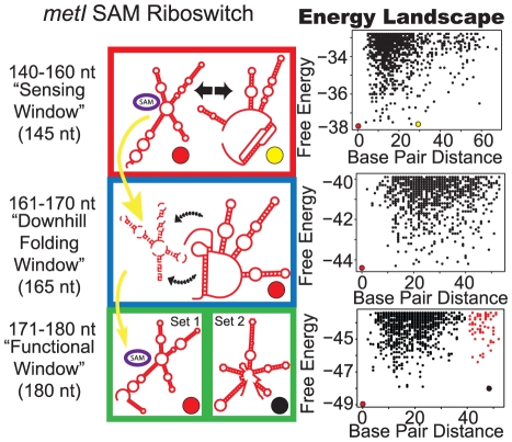
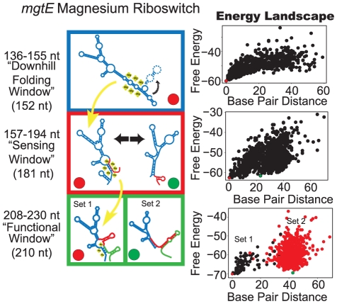
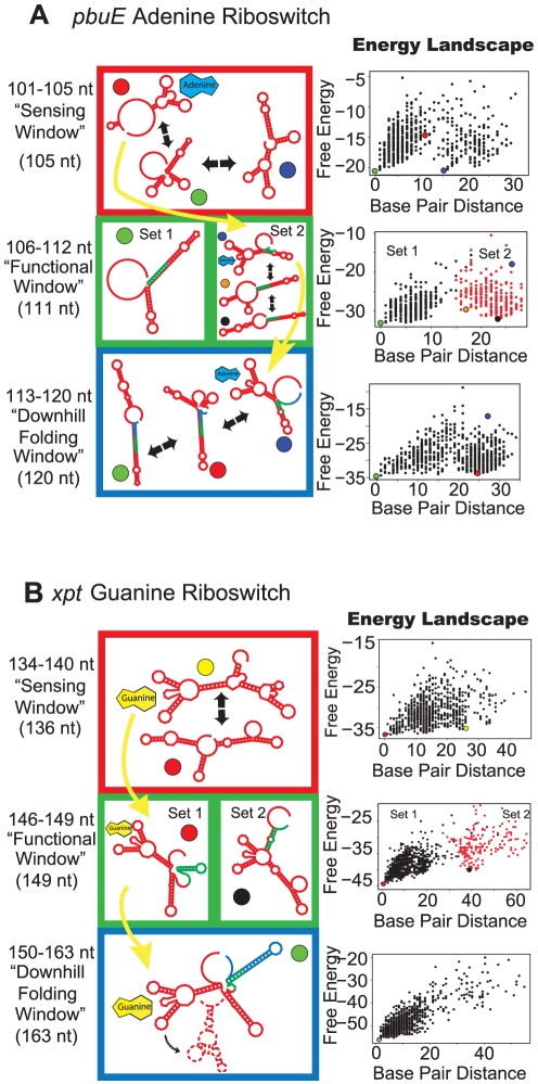
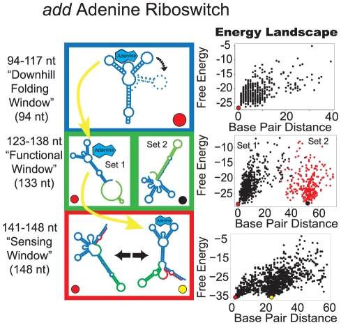
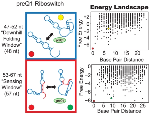
Riboswitches are RNAs that modulate gene expression by ligand-induced conformational changes. However, the way in which sequence dictates alternative folding pathways of gene regulation remains unclear. In this study, we compute energy landscapes, which describe the accessible secondary structures for a range of sequence lengths, to analyze the transcriptional process as a given sequence elongates to full length. In line with experimental evidence, we find that most riboswitch landscapes can be characterized by three broad classes as a function of sequence length in terms of the distribution and barrier type of the conformational clusters: low-barrier landscape with an ensemble of different conformations in equilibrium before encountering a substrate; barrier-free landscape in which a direct, dominant "downhill" pathway to the minimum free energy structure is apparent; and a barrier-dominated landscape with two isolated conformational states, each associated with a different biological function. Sharing concepts with the "new view" of protein folding energy landscapes, we term the three sequence ranges above as the sensing, downhill folding, and functional windows, respectively. We find that these energy landscape patterns are conserved in various riboswitch classes, though the order of the windows may vary. In fact, the order of the three windows suggests either kinetic or thermodynamic control of ligand binding. These findings help understand riboswitch structure/function relationships and open new avenues to riboswitch design.


Conflict of interest statement

The authors have declared that no competing interests exist.

Figures

Figure 1
Figure 1. The riboswitch control of gene regulation.
(a) The two full length structures of the tenA thiamine pyrophosphate (TPP, blue oval) riboswitch are shown. Aptamer domain is highlighted in orange solid and broken lines. (b) Simplified diagram of riboswitch folding process for the tenA TPP riboswitch. From 156–171 nt, meta-stable structures (labeled ligand-competent and non-ligand-competent) exchange with one another. From 172–179 nt, ligand (yellow polygon) stabilizes one of the ligand-competent meta-stable structures, thus causing the formation of specific terminator hairpin structures in the full length riboswitch (181–190 nt). The ligand may remain bound or disengage the aptamer later in transcription (polygon in dashed lines in top row, right column). In the absence of ligand (172–179 nt), isomerization to a different structure occurs, causing an antiterminator structure to form downstream (181–190 nt). (c) The energy landscapes of the riboswitch through all three stages of transcription. Each point represents a different secondary structure; marked according to its base pair distance (structure distance) and free energy. Colored stars represent points on the landscape corresponding to structures in (1b). In the energy landscape from 156–171 nt, represented here by the landscape at 165 nt, a small energy barrier and multiple low-energy structures exist on the landscape simultaneously, permitting exchange of meta-stable structures (double arrow). From 172–179 nt (shown for 175 nt), the landscapes are funnel-shaped and cause isomerization to the mfe if ligand is not present to stabilize the ligand-competent structure. From 181–190 nt (shown here for 185 nt), two structures are possible which the RNA may fold into. The high energy barrier between sets precludes switching. Set 1 corresponds to terminator structures and Set 2 corresponds to antiterminator structures. (d) Example calculation of base-pair distance for a simple helix structure.
Figure 2
Figure 2. Proposed folding pathway for the TPP riboswitches tenA (a) and thiM (b).
Structures formed in the sensing windows are represented in red boxes; downhill folding window structures are found in blue boxes; and functional window structures are represented inside the green boxes. Double-head arrows represent structures that can interchange. Broken-line structural elements in downhill folding window (blue box) represent structural elements that would be coerced to form in the presence of ligand. Colored circles adjacent to structures are marked by their points on the respective energy landscape to the right. Yellow arrows represent the series of structures accessed in the presence of ligand. For all sequence lengths inside of a window, the energy landscape repeatedly displays similar patterns (see Materials and Methods). The specific sequence length corresponding to the window shown is given following the length range. For full description of energy landscape characteristics see Figure S5.
Figure 3
Figure 3. Proposed folding pathways for the GEMM (a) and moaA (b) riboswitches.
See figure 2 caption for description of figure elements. For full description of energy landscape characteristics see Figure S5.
Figure 4
Figure 4. Proposed folding pathway for the S-adenosylmethionine (SAM) metI riboswitch.
See figure 2 caption for description of figure elements. For full description of energy landscape characteristics see Figure S5.
Figure 5
Figure 5. Proposed folding pathway for the mgtE riboswitch.
See figure 2 caption for description of figure elements. For full description of energy landscape characteristics see Figure S5.
Figure 6
Figure 6. Proposed folding pathway for the pbuE (a) and xpt (b) purine riboswitches.
See figure 2 caption for description of figure elements. For full description of energy landscape characteristics see Figure S5.
Figure 7
Figure 7. Proposed folding pathway for the add (a) riboswitch.
See figure 2 caption for description of figure elements. For full description of energy landscape characteristics see Figure S5.
Figure 8
Figure 8. Proposed folding pathway for the preQ1 riboswitch.
See figure 2 caption for description of figure elements. For full description of energy landscape characteristics see Figure S5.

Similar articles

Cited by

References

    1. Barrick JE, Breaker RR. The distributions, mechanisms, and structures of metabolite-binding riboswitches. Genome Biol. 2007;8:R239. - PMC - PubMed
    1. Montange RK, Batey RT. Riboswitches: emerging themes in RNA structure and function. Annu Rev Biophys. 2008;37:117–133. - PubMed
    1. Smith AM, Fuchs RT, Grundy FJ, Henkin TM. Riboswitch RNAs: regulation of gene expression by direct monitoring of a physiological signal. RNA Biol. 2010;7:104–110. - PMC - PubMed
    1. Wachter A. Riboswitch-mediated control of gene expression in eukaryotes. RNA Biol. 2010;7:67–76. - PubMed
    1. Epshtein V, Mironov AS, Nudler E. The riboswitch-mediated control of sulfur metabolism in bacteria. Proc Natl Acad Sci U S A. 2003;100:5052–5056. - PMC - PubMed

Publication types