. 2012 Feb;8(2):e1002532.
doi: 10.1371/journal.ppat.1002532. Epub 2012 Feb 16.

Borrelia burgdorferi requires the alternative sigma factor RpoS for dissemination within the vector during tick-to-mammal transmission

Affiliations

Borrelia burgdorferi requires the alternative sigma factor RpoS for dissemination within the vector during tick-to-mammal transmission

Star M Dunham-Ems et al. PLoS Pathog. 2012 Feb.

Abstract

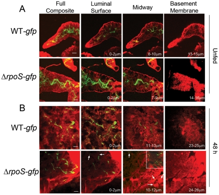
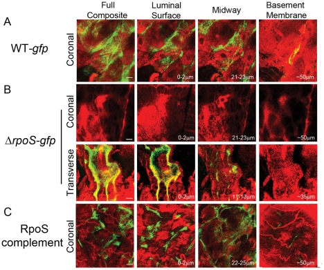
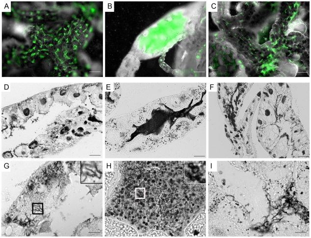
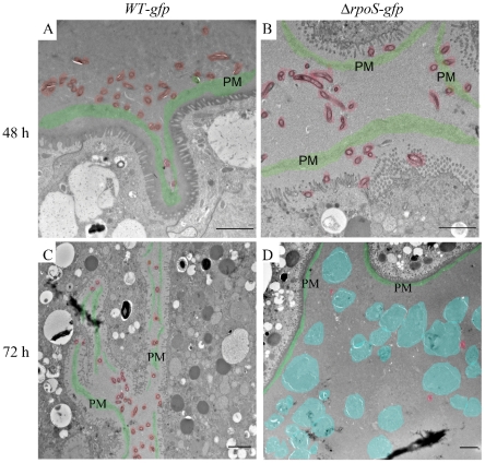
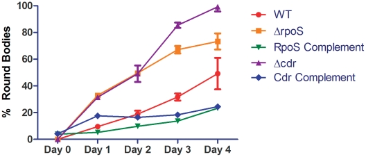
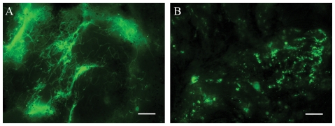
While the roles of rpoS(Bb) and RpoS-dependent genes have been studied extensively within the mammal, the contribution of the RpoS regulon to the tick-phase of the Borrelia burgdorferi enzootic cycle has not been examined. Herein, we demonstrate that RpoS-dependent gene expression is prerequisite for the transmission of spirochetes by feeding nymphs. RpoS-deficient organisms are confined to the midgut lumen where they transform into an unusual morphotype (round bodies) during the later stages of the blood meal. We show that round body formation is rapidly reversible, and in vitro appears to be attributable, in part, to reduced levels of Coenzyme A disulfide reductase, which among other functions, provides NAD+ for glycolysis. Our data suggest that spirochetes default to an RpoS-independent program for round body formation upon sensing that the energetics for transmission are unfavorable.


Conflict of interest statement

The authors have declared that no competing interests exist.

Figures

Figure 1
Figure 1. Contours of the RpoSBb regulon in I. scapularis.
qRT-PCR analysis of (A) absolutely and (B) partially RpoS-dependent upregulated genes and (C) RpoS-repressed genes selected from microarray data derived from Bb cultivated within DMCs . A representative sample of genes is shown; data for the remaining genes are presented in Figure S2. Expression profiling was performed using fed larvae, unfed and fed nymphs that had been naturally-infected with WT Bb as well as fed nymphs that had been infected as larvae by immersion with either WT-gfp or ΔrpoS-gfp isolates. Values represent the average flaB-normalized transcript copy number ± standard error of the mean (SEM) for each gene; values are considered significantly different when p is ≤0.05 (indicated by asterisks). Composite confocal images through the full thickness of nymphal midguts at 72 h post-placement showing the distribution of spirochetes expressing gfp under the control of the (D) flaB or (E) ospA promoter. A detailed schematic indicating how confocal images of fed midguts were acquired is presented in Figure S3. Here and elsewhere, green represents GFP+ spirochetes while red indicates midgut epithelial cells labeled with FM4-64; scale bars = 25 µm.
Figure 2
Figure 2. Spirochetes lacking RpoS are distributed normally within unfed nymphal midguts but are destroyed between epithelial cells early during feeding.
The leftmost images in each panel depict the full composites (basement membrane to basement membrane) of the midgut, while 3-µm composite images show spirochetes at the luminal surface, midway through the epithelium, and at the basement membrane; scale bars = 25 µm. A detailed schematic indicating how confocal images of unfed and 48 h-fed midguts were acquired is presented in Figure S3. (A) In unfed midguts, WT-gfp and ΔrpoS-gfp are similarly distributed. (B) In midguts isolated at 48 h post-placement, WT-gfp organisms form aggregates, while ΔrpoS-gfp located between epithelial cells are destroyed; arrows indicate ragged, blebbing Bb. Numbers in lower right hand corner indicate the optical depth of the image. Inset in the ΔrpoS-gfp midway panel depicts a 1-µm optical section showing thin, ragged and blebbing organisms between epithelial cells; see also Figure S4 for a comparison of midguts infected with WT-gfp Bb by natural versus immersion methods.
Figure 3
Figure 3. RpoS is required to form networks during nymphal feeding.
(A) At 72 h post-placement, WT-gfp organisms form networks that encase midgut epithelial cells and extend to the basement membrane. (B) ΔrpoS-gfp Bb are not observed in intact midguts imaged in the ‘coronal’ plane but are visualized within the lumens of midguts that had been punctured to remove a portion of their contents and imaged by acquiring transverse optical sections from distal portions of the diverticula to reduce light scattering and absorption by the blood meal. A detailed schematic illustrating how coronal and transverse confocal images were acquired is presented in Figure S3. (C) Complementation restores the ability of ΔrpoS-gfp Bb to form networks and extend to the basement membrane; see also Figure S5 for complementation verification by protein profile analysis. The leftmost images in each panel depict the full composites (basement membrane to luminal surface) of the midgut, while 3-µm composite images show spirochetes at the luminal surface, midway through the epithelial layer, and at the basement membrane. Numbers in lower right hand corner indicate the optical depth of the image; scale bars = 25 µm. Representative cryosections of 72 h-fed midguts infected with WT-gfp and ΔrpoS-gfp isolates are presented in Figure S6.
Figure 4
Figure 4. Spirochetes lacking RpoS are confined to the lumen and develop into round bodies during the later stages of feeding.
(A–C) Representative epifluorescence images of midguts isolated at 72 h post-placement containing (A) WT-gfp, (B) ΔrpoS-gfp or (C) RpoS-complemented isolates; midguts appear white when imaged by dark-field microscopy. (D–I) Representative silver-stained paraffin-embedded sections of midguts isolated from 72 h-fed nymphs infected with (D and G) WT-gfp, (E and H) ΔrpoS-gfp or (F and I) RpoS-complemented isolates. Insets in (G) and (H) are enlargements of the boxed regions showing WT-gfp spirochetes and ΔrpoS-gfp round bodies, respectively; scale bars: A–C = 50 µm; D–F = 25 µm; and G–I = 10 µm.
Figure 5
Figure 5. ΔrpoS Bb form round bodies within nymphal midguts during the later stages of feeding.
Representative TEM images of nymphal midguts containing WT-gfp and ΔrpoS-gfp spirochetes isolated at (A–B) 48 or (C-D) 72 h post-placement. Color overlays are used to highlight normal spirochetes (red), round bodies (blue), and peritrophic membranes (PM, green); scale bars = 2 µm.
Figure 6
Figure 6. Wild-type and ΔrpoS round bodies are structurally similar both within feeding nymphs and under nutrient-limiting conditions in vitro.
Representative TEM images of ΔrpoS-gfp and WT-gfp spirochetes and round bodies in (A) nymphal midguts isolated at 48 or 72 h post-placement or (B) in vitro-derived organisms following incubation in BSK-II or RPMI for 3 days. In panels A and B, arrows indicate examples of protoplasmic cylinders; scale bars = 500 nm.
Figure 7
Figure 7. Loss of RpoS and CoADR exacerbates round body formation in vitro.
Bb were incubated in RPMI for 1–4 days. A minimum of 300 organisms were counted per strain for each time point. Experiments were performed in triplicate; error bars represent means ± SEM. Representative images of fields used to quantify round body formation are shown in Figure S7. The percentages of round bodies formed by ΔrpoS-gfp and Δcdr isolates were significantly greater than WT on days 1 through 4 (p≤0.002). Round body formation by ΔrpoS-gfp and Δcdr isolates was significantly different on days 3 and 4 (p≤0.002).
Figure 8
Figure 8. Round bodies within fed nymphal midguts recover into elongated spirochetes.
ΔrpoS-infected nymphs were removed at 72 h post-placement and midguts dissected into RPMI. (A) The addition of BSK-II induced the recovery of round bodies into elongated spirochetes. (B) Round bodies do not recover when midguts were submerged in RPMI. Scale bars = 10 µm. See also the Video S1 of the rapid recovery of in vitro-derived organisms.

Similar articles

Cited by

References

    1. Gruber TM, Gross CA. Multiple sigma subunits and the partitioning of bacterial transcription space. Annu Rev Microbiol. 2003;57:441–466. - PubMed
    1. Hengge-Aronis R. Recent insights into the general stress response regulatory network in Escherichia coli. J Mol Microbiol Biotechnol. 2002;4:341–346. - PubMed
    1. Battesti A, Majdalani N, Gottesman S. The RpoS-Mediated General Stress Response in Escherichia coli. Annu Rev Microbiol. 2011;65:189–213. - PMC - PubMed
    1. Hengge-Aronis R. Signal transduction and regulatory mechanisms involved in control of the σs (RpoS) subunit of RNA polymerase. Microbiol Mol Biol Rev. 2002;66:373–395. - PMC - PubMed
    1. Klauck E, Typas A, Hengge R. The σS subunit of RNA polymerase as a signal integrator and network master regulator in the general stress response in Escherichia coli. Sci Prog. 2007;90:103–127. - PMC - PubMed

Publication types