. 2012;8(2):e1002519.
doi: 10.1371/journal.pgen.1002519. Epub 2012 Feb 16.

Repression of a potassium channel by nuclear hormone receptor and TGF-β signaling modulates insulin signaling in Caenorhabditis elegans

Affiliations

Repression of a potassium channel by nuclear hormone receptor and TGF-β signaling modulates insulin signaling in Caenorhabditis elegans

Donha Park et al. PLoS Genet. 2012.

Abstract

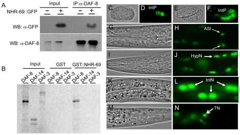
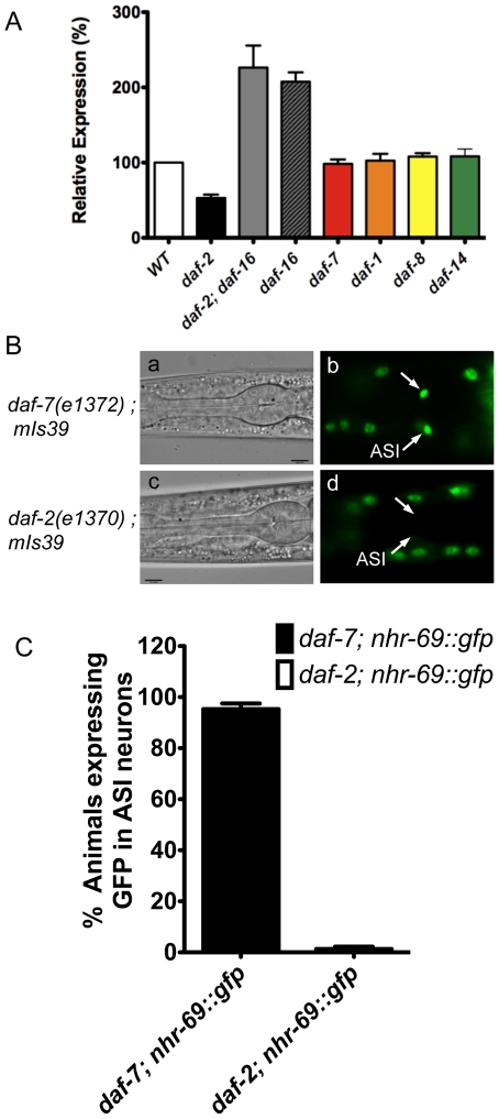
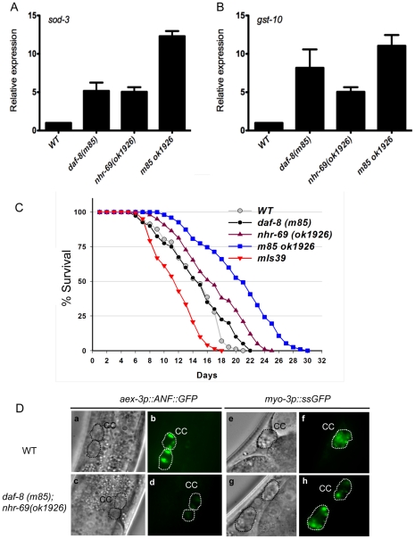
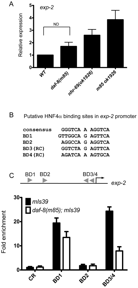
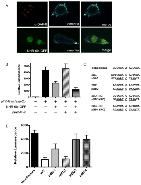
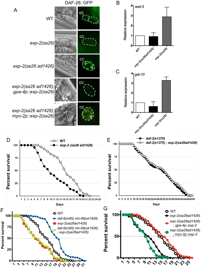
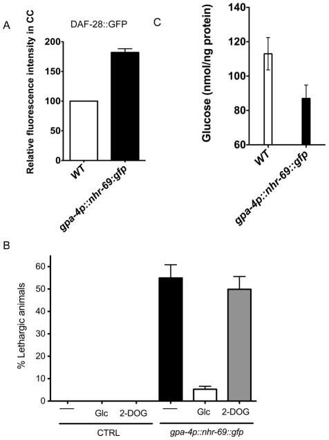
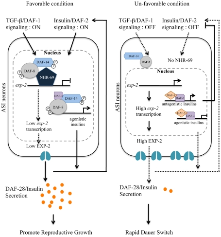
Transforming growth factor β (TGF-β) signaling acts through Smad proteins to play fundamental roles in cell proliferation, differentiation, apoptosis, and metabolism. The Receptor associated Smads (R-Smads) interact with DNA and other nuclear proteins to regulate target gene transcription. Here, we demonstrate that the Caenorhabditis elegans R-Smad DAF-8 partners with the nuclear hormone receptor NHR-69, a C. elegans ortholog of mammalian hepatocyte nuclear factor 4α HNF4α), to repress the exp-2 potassium channel gene and increase insulin secretion. We find that NHR-69 associates with DAF-8 both in vivo and in vitro. Functionally, daf-8 nhr-69 double mutants show defects in neuropeptide secretion and phenotypes consistent with reduced insulin signaling such as increased expression of the sod-3 and gst-10 genes and a longer life span. Expression of the exp-2 gene, encoding a voltage-gated potassium channel, is synergistically increased in daf-8 nhr-69 mutants compared to single mutants and wild-type worms. In turn, exp-2 acts selectively in the ASI neurons to repress the secretion of the insulin-like peptide DAF-28. Importantly, exp-2 mutation shortens the long life span of daf-8 nhr-69 double mutants, demonstrating that exp-2 is required downstream of DAF-8 and NHR-69. Finally, animals over-expressing NHR-69 specifically in DAF-28-secreting ASI neurons exhibit a lethargic, hypoglycemic phenotype that is rescued by exogenous glucose. We propose a model whereby DAF-8/R-Smad and NHR-69 negatively regulate the transcription of exp-2 to promote neuronal DAF-28 secretion, thus demonstrating a physiological crosstalk between TGF-β and HNF4α-like signaling in C. elegans. NHR-69 and DAF-8 dependent regulation of exp-2 and DAF-28 also provides a novel molecular mechanism that contributes to the previously recognized link between insulin and TGF-β signaling in C. elegans.


Conflict of interest statement

The authors have declared that no competing interests exist.

Figures

Figure 1
Figure 1. Interaction of NHR-69 with DAF-8 and expression pattern of nhr-69p::nhr-69::gfp.
(A) In vivo co-immunoprecipitation was performed with anti-DAF-8 antibody from wild-type worms and nhr-69p::nhr-69::gfp expressing animals, followed by immunoblot with anti-GFP antibody to detect NHR-69::GFP. (B) In vitro GST pull-down assay between NHR-69 and SMAD proteins (DAF-8, DAF-14, or DAF-3). C–N show DIC and fluorescence images of developing embryos (C–F) and of adults (G–N) expressing nhr-69p::nhr-69::gfp. (C and D) Expression at the E16 stage of intestinal precursor cells in embryos. (E and F) Expression at the comma stage. (G and H) Expression in ASI neurons (arrows). (I and J) Hypodermal expression. (K and L) Intestinal expression. (M and N) Expression in tail neurons. IntP; Intestinal precursor cells, HypN; Hypodermal nucleus, IntN; Intestinal nucleus, TN; Tail neurons.
Figure 2
Figure 2. Insulin signaling positively regulates nhr-69 expression.
(A) The bars represent relative levels of nhr-69 mRNA as determined by qPCR on RNA isolated from daf-2(e1370), daf-2(e1370); daf-16(mgDf47), daf-16(mgDf47), daf-1(m40), daf-7(e1372), daf-8(m85) and daf-14(m77) mutants (relative to mRNA levels in wild-type worms); mRNA levels were normalized to act-2. Error bars indicate standard errors of the mean (SEM) from four biological replicates. (B) Micrographs show DIC and fluorescence images of NHR-69::GFP in daf-7(e1372) (a and b) and daf-2(e1370) (c and d) mutants. Arrows indicate the ASI neurons. All fluorescence images were taken with identical exposure times. (C) Bar graphs indicate the percentage of animals expressing NHR-69::GFP in ASI neurons in daf-7(e1372) (black bar, N = 78) and in daf-2(e1370) mutant (white bar, N = 83) shown in (B). Error bars indicate SEM.
Figure 3
Figure 3. Reduced insulin signaling and neuropeptide secretion in daf-8 nhr-69 mutants.
(A and B) Bar graphs show the relative mRNA levels of sod-3 and gst-10 in daf-8(m85), nhr-69(ok1926) and daf-8(m85) nhr-69(ok1926) mutants relative to wild-type worms, as determined by qPCR (normalized to act-2). Age synchronized day-1 adults were used for total RNA extraction. Error bars indicate SEM from three biological replicates. (C) The plots show population survival of wild-type (grey circles), daf-8(m85) (black circles), nhr-69(ok1926) (purple triangles), daf-8(m85) nhr-69(ok1926) (blue squares), and mIs39[nhr-69p::nhr-69::gfp] (red inverted triangles) worms at 25.5°C. (D) DIC and fluorescence micrographs show coelomocyte accumulation (CC, dotted circles) of ANF::GFP (expressed from the oxIs206[aex-3p::ANF::gfp] transgene; b and d) or ssGFP (expressed from the arIs37[myo-3P::ssGFP] transgene; f and h), respectively, in wild-type worms and in daf-8(m85) nhr-69(ok1926) mutants, as indicated.
Figure 4
Figure 4. NHR-69 directly regulates exp-2 transcription.
(A) The bar graphs represent the relative levels of exp-2 mRNA in daf-89(m85), nhr-69(ok1926), and daf-8(m85) nhr-69(ok1926) mutants relative to wild type as determined by qPCR (normalized to act-2). Error bars indicate SEM from three biological replicates. Age synchronized day-1 adults were used for total RNA extraction. (B) HNF4α binding consensus sequence and putative NHR-69/HNF4α-like binding sites in the exp-2 promoter region. The positions of possible binding sites 5′ of the ATG of exp-2 are BD1 (−2342 bp), BD2 (−1832 bp), BD3 (−118 bp) and BD4 (−86 bp), respectively. RC, reverse complement. (C) ChIP-qPCR with anti-GFP antibody reveals that NHR-69 is directly associated with at least two sites in the 5′ regulatory region of exp-2. The arrowheads indicate the putative HNF4α binding sites shown in (B). Anti-GFP antibody precipitated sequences from BD1 and BD3 and/or BD4. In a daf-8 background, binding of NHR-69::GFP was reduced 27% for BD1 and 71% for BD3/BD4. Error bars indicate SEM from three biological replicates.
Figure 5
Figure 5. NHR-69 and DAF-8 suppress exp-2 promoter activity in mammalian cells.
(A) Plasmids expressing NHR-69::GFP and a phosphomimetic variant of DAF-8 (pmDAF-8) were transiently transfected into HEK293 cells and visualized by fluorescence imaging; both proteins show nuclear localization. (B) The bar graphs depict relative luciferase activity resulting from a transiently transfected exp-2-driven luciferase reporter in HEK293 cells. NHR-69, but not pmDAF-8 single transfection represses the exp-2 promoter, and cotransfection of NHR-69 and pmDAF-8 causes an even stronger repression. (C) Details on the site-directed mutagenesis of HNF4α-like binding sites in the exp-2 promoter. (D) The graph depicts the consequence of site directed mutagenesis of HNF4α-like binding sites in the exp-2 promoter on luciferase activity. “No effectors” indicates transfection of the reporter alone, whereas “WT” indicates co-transfection of NHR-69 and pmDAF-8 with the wild-type exp-2 promoter (similar to panel B). Mutating BD1, BD3, or BD4 attenuates the NHR-69 and pmDAF-8-mediated repression of exp-2 activity. The result is an average from three individual trials.
Figure 6
Figure 6. Insulin signaling is impaired in exp-2 mutants.
(A) The micrographs show DAF-28::GFP accumulation in coelomocytes (CC) in wild-type worms, in exp-2 mutants, and in exp-2 transgenic strains. exp-2(sa26ad1426) loss-of-function and exp-2(sa26) gain-of-function mutants show increased and decreased DAF-28::GFP intensity, respectively, indicating that EXP-2 represses DAF-28 secretion. ASI-neuron specific expression (driven by the gpa-4 promoter) of the gain-of-function exp-2(sa26) allele in the exp-2(sa26ad1426) background reduces DAF-28::GFP accumulation, resulting in a level similar to that seen in the global exp-2(sa26) gain-of-function mutant; in contrast, pharyngeal-specific exp-2(sa26) expression (driven by the myo-2 promoter) does not alter DAF-28::GFP accumulation in the exp-2(sa26ad1426) background. (B and C) The bar graphs depict average sod-3 and gst-10 mRNA levels in wild-type (white), exp-2(sa26ad1426) (black), and exp-2(sa26) (grey) worms, as determined by qPCR (normalized to act-2). Error bars indicate SEM from four biological replicates. (D) The graph depicts population survival curves for wild type worms (white) and exp-2(sa26ad1426) loss-of-function mutants (black; T = 25.5°C). (E) The graph shows population survival curves of daf-2(e1370) (black) and daf-2(e1370); exp-2(sa26ad1426) mutants (white; T = 25.5°C). (F) The graph depicts population survival curves of wild-type (black circle), daf-8(m85) nhr-69(ok1926) (blue circle), exp-2(sa26ad1426) (red square), and daf-8(m85) nhr-69(ok19260; exp-2(sa26ad1426) (green square) worms (T = 25.5°C). (G) The graph shows population survival curves of wild-type (white), exp-2(sa26ad1426) (black), exp-2(sa26ad1426); gpa-4p::exp-2 (red) and exp-2(sa26ad1426); myo-2p::exp-2 (green) (T = 25.5°C). All strains shown in this panel also harbor the pRF4[rol-6(su1006)] transgene.
Figure 7
Figure 7. ASI-specific over-expression of NHR-69 confers hypoglycemia.
(A) The bar graphs show the relative DAF-28::GFP level in coelomocytes (CC) in wild-type and mIs40[gpa-4p::nhr-69::gfp] day-1 old adult worms. Error bars indicate SEM. N = 62 in wild type, N = 77 in mIs40[gpa-4p::nhr-69::gfp] backgrounds (p<0.001). (B) Bars indicate the percentage of lethargic animals in the presence or absence of exogenous glucose (2 mM Glc) or 2-dexoyglucose (2 mM 2-DOG). Synchronous day-1 adults were video recorded for two minutes individually and worms that stopped and resumed moving more than four times were judged to be lethargic (N = 50). (C) Bars show endogenous glucose content in wild-type and mIs40[gpa-4p::nhr-69::gfp] worms. Synchronous day-1 adults were assayed in four independent biological replicates. Error bars indicate SEM.
Figure 8
Figure 8. Working model for DAF-8 and NHR-69 modulation of insulin secretion in C. elegans.
Based on our data, we propose that DAF-2 signaling positively regulates transcription of nhr-69, whereas the TGF-β receptor is known to phosphorylate DAF-8 to activate its function . NHR-69 and DAF-8 cooperatively repress the transcription of the exp-2 Kv channel gene in ASI neurons, which in turn causes sustained secretion of the insulin-like peptide DAF-28, hence activating a feed-forward loop on DAF-2 in favorable conditions. TGF-β and Insulin/IGF-1 signaling crosstalk at the exp-2 promoter through the DAF-8 and NHR-69 transcription factors. Upregulation of exp-2 by the reduction of DAF-8 and NHR-69 activity would attenuate DAF-28 secretion, providing a fast decision for the dauer formation.

Similar articles

Cited by

References

    1. Massagué J, Gomis R. The logic of TGFbeta signaling. FEBS Lett. 2006;580:2811–20. - PubMed
    1. Attisano L, Carcamo J, Ventura F, Weis F. Identification of Human Activin and TGF Type I Receptors That Form Heteromeric Kinase Complexes. CELL-CAMBRIDGE MA- 1993;75:671–680. - PubMed
    1. Savage C, Das P, Finelli A, Townsend S, Sun C, et al. Caenorhabditis elegans genes sma-2, sma-3, and sma-4 define a conserved family of transforming growth factor beta pathway components. Proc Natl Acad Sci USA. 1996;93:790–4. - PMC - PubMed
    1. Riddle DL, Swanson MM, Albert PS. Interacting genes in nematode dauer larva formation. Nature. 1981;290:668–671. - PubMed
    1. Hu P. Dauer. WormBook: the online review of C elegans biology. 2007:1–19. - PMC - PubMed

Publication types

MeSH terms