. 2012;8(2):e1002528.
doi: 10.1371/journal.pgen.1002528. Epub 2012 Feb 16.

Discovery of a modified tetrapolar sexual cycle in Cryptococcus amylolentus and the evolution of MAT in the Cryptococcus species complex

Affiliations

Discovery of a modified tetrapolar sexual cycle in Cryptococcus amylolentus and the evolution of MAT in the Cryptococcus species complex

Keisha Findley et al. PLoS Genet. 2012.

Abstract

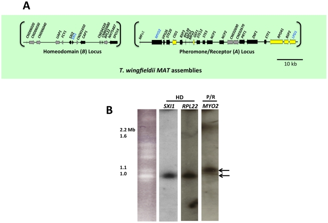
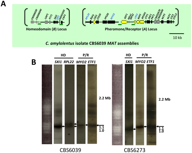
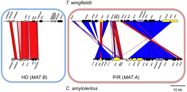
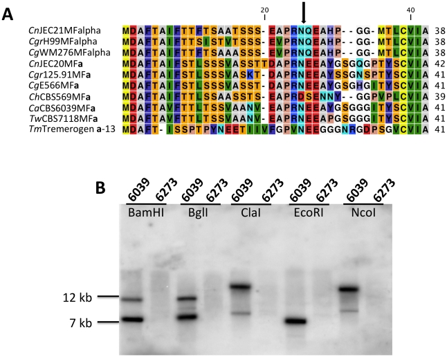
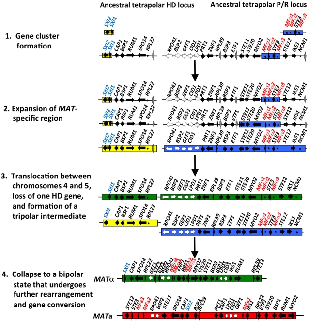
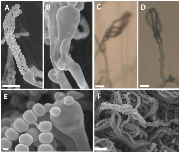
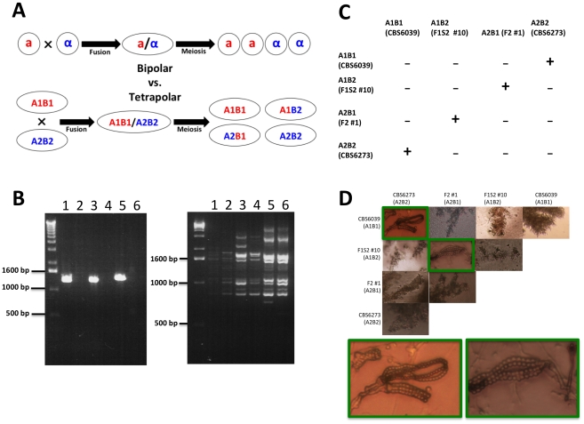
Sexual reproduction in fungi is governed by a specialized genomic region called the mating-type locus (MAT). The human fungal pathogenic and basidiomycetous yeast Cryptococcus neoformans has evolved a bipolar mating system (a, α) in which the MAT locus is unusually large (>100 kb) and encodes >20 genes including homeodomain (HD) and pheromone/receptor (P/R) genes. To understand how this unique bipolar mating system evolved, we investigated MAT in the closely related species Tsuchiyaea wingfieldii and Cryptococcus amylolentus and discovered two physically unlinked loci encoding the HD and P/R genes. Interestingly, the HD (B) locus sex-specific region is restricted (∼2 kb) and encodes two linked and divergently oriented homeodomain genes in contrast to the solo HD genes (SXI1α, SXI2a) of C. neoformans and Cryptococcus gattii. The P/R (A) locus contains the pheromone and pheromone receptor genes but has expanded considerably compared to other outgroup species (Cryptococcus heveanensis) and is linked to many of the genes also found in the MAT locus of the pathogenic Cryptococcus species. Our discovery of a heterothallic sexual cycle for C. amylolentus allowed us to establish the biological roles of the sex-determining regions. Matings between two strains of opposite mating-types (A1B1×A2B2) produced dikaryotic hyphae with fused clamp connections, basidia, and basidiospores. Genotyping progeny using markers linked and unlinked to MAT revealed that meiosis and uniparental mitochondrial inheritance occur during the sexual cycle of C. amylolentus. The sexual cycle is tetrapolar and produces fertile progeny of four mating-types (A1B1, A1B2, A2B1, and A2B2), but a high proportion of progeny are infertile, and fertility is biased towards one parental mating-type (A1B1). Our studies reveal insights into the plasticity and transitions in both mechanisms of sex determination (bipolar versus tetrapolar) and sexual reproduction (outcrossing versus inbreeding) with implications for similar evolutionary transitions and processes in fungi, plants, and animals.


Conflict of interest statement

The authors have declared that no competing interests exist.

Figures

Figure 1
Figure 1. T. wingfieldii MAT loci and chromosomal locations.
(A) Six fosmids were analyzed to generate the assembly for T. wingfieldii. The MAT gene probes used to probe the T. wingfieldii library are indicated in blue. The HD (B) and P/R (A) loci are embedded within assemblies that span 40 and 70 kb respectively. Grey arrows indicate genes that either flank MAT or are hypothetical genes, black arrows are Cryptococcus MAT-specific genes, and yellow indicates the genes most recently acquired into the Cryptococcus MAT locus. Scale bar = 10 kb. (B) Chromosomes from T. wingfieldii were separated using PFGE, followed by Southern hybridization using three MAT-specific probes, two from the HD locus and one from the P/R locus. Arrows depict hybridization of HD genes to an ∼1 Mb chromosome distinct from hybridization of the P/R genes to an ∼1.1 Mb chromosome.
Figure 2
Figure 2. C. amylolentus MAT loci and chromosomal locations.
(A) Four fosmids were analyzed to generate the assembly for C. amylolentus. The MAT gene probes used to probe the C. amylolentus library are indicated in blue. The HD (B) and P/R (A) loci are embedded in regions that span 20 and 60 kb respectively. Grey arrows indicate genes that either flank MAT or are hypothetical genes, black arrows are Cryptococcus MAT-specific genes, and yellow indicates the genes more recently acquired into the Cryptococcus MAT locus. Several gaps remain in the MAT loci of C. amylolentus. Scale bar = 10 kb. Green bars under the assembly denote gaps in sequence. (B) Chromosomes from C. amylolentus were separated using PFGE, and analyzed by Southern hybridization using three MAT-specific probes, one from the HD locus and two from the P/R locus. The RPL22 gene was also used as a probe. Arrows depict hybridization of the HD and P/R locus probes to distinct chromosomes (∼1.1 and ∼1.15 Mb).
Figure 3
Figure 3. Synteny analysis of MAT sequences from T. wingfieldii and C. amylolentus.
On the left is the comparison of the HD (MAT B) locus, while the comparison of the P/R (MAT A) locus is shown on the right. Red lines connecting T. wingfieldii and C. amylolentus sequences denote conserved gene order; while blue lines indicate inverted orientations of the sequences from the two species. Green bars under the assembly denote sequence gaps in the assembled contigs.
Figure 4
Figure 4. Analysis of the pheromone/receptor genes in C. amylolentus.
(A) Sequence alignments of the pheromone gene in C. neoformans var. neoformans JEC21 MFα, C. neoformans var. grubii H99 MFα, C. gattii WM276 MFα, C. neoformans var. neoformans JEC20 MF a, C. neoformans var. grubii 125.91 MF a, C. gattii E566 MF a, C. heveanensis CBS569 MF a, C. amylolentus CBS6039 MF a1, T. wingfieldii CBS7118 MF a1, and T. mesenterica ATCC24925 Tremerogen a-13. The black arrow denotes the predicted cleavage site. The pheromone receptor gene, STE3, is MAT specific. (B) Genomic DNA from the two C. amylolentus strains was digested with BamHI, BglI, ClaI, EcoRI, or NcoI and Southern blot analysis was performed using the STE3 PCR product from CBS6039 as a probe. For each enzyme digestion, CBS6039 was in the left lane and CBS6273 was in the right lane.
Figure 5
Figure 5. Model for the evolution of the mating-type locus in the pathogenic Cryptococcus species.
The physically unlinked ancestral tetrapolar HD and P/R loci contained both homeodomain genes and the pheromone/receptor genes respectively. Additional genes were acquired into both loci, expanding the MAT-specific region. A translocation event occurred, likely between chromosomes 4 and 5 of Cryptococcus, resulting in the formation of a transient tripolar intermediate and one of the HD genes was lost. The hypothetical genes (grey arrows) relocated, likely through a translocation event, to the telomeric ends of chromosome 4. The unstable tripolar intermediate later collapsed to a bipolar state. The fused loci were subjected to further gene rearrangement and gene conversion events, which led to the formation of the bipolar alleles of the pathogenic Cryptococcus species. White arrows indicate the five genes most recently acquired into Cryptococcus MAT locus and black arrows are MAT-specific genes present in the pathogenic Cryptococcus species.
Figure 6
Figure 6. The homeodomain genes, SXI1 and SXI2, define MAT.
A percent identity plot of both C. amylolentus strains, CBS6039 and CBS6273, comparing the SXI1 and SXI2 dimorphic region in the HD locus. The red ellipsoid represents an EcoRV site, which only cleaves SXI1 in CBS6039 while the blue ellipsoid represents an RsaI site, which only cleaves SXI2 in CBS6039.
Figure 7
Figure 7. Phylogenetic patterns of four C. amylolentus MAT genes.
The phylogenetic relationships of C. amylolentus to the pathogenic Cryptococcus species and neighboring taxa based on four genes, GEF1, CID1, SXI1, and SXI2, are shown. GEF1 and CID1 display a species-specific phylogeny and the SXI1 and SXI2 alleles are very diverged from the pathogenic Cryptococcus species. The trees were constructed using the Neighbor-Joining method implemented in the software MEGA4. Bootstrap values on tree branches were calculated from 500 replicates. (α) indicates strains with the MATα locus, and (a) indicates strains with the MAT a locus.
Figure 8
Figure 8. Sexual reproduction of C. amylolentus.
Microscopic examination of mating structures produced during sex between the two C. amylolentus strains, CBS6039 and CBS6273, on V8 (pH = 5) medium incubated in the dark at room temperature for 2 weeks. (A) SEM of basidiospores attached to basidia. Scale bar represents 10 µm. (B) SEM of fused and unfused clamp connections. (C and D) Light microscopy at a magnification of 20X of hyphal filaments, basidia, and basidiospores, scale bar = 10 µm. (E) Basidium with youngest spores attached and associated detached spore chains, scale bar = 1 µm. (F) A cluster of basidiospores and basidia. Scale bar = 10 µm.
Figure 9
Figure 9. C. amylolentus has a tetrapolar mating system.
(A) In a bipolar mating system, haploid a and α cells fuse to form a diploid a/α cell. Sex culminates in meiosis, which gives rise to four meiotic progeny, 2 a and 2 α. The a progeny can mate with the α parent (50%) while the α progeny can mate with the a parent (50%). In a tetrapolar mating system, haploid A1B1 and A2B2 cells fuse to form a dikaryon/diploid A1B1/A2B2. Meiosis then results in the production of four haploid meiotic progeny: A1B1 can mate with the A2B2 parent and progeny (25%), A2B2 can mate with the A1B1 parent and progeny (25%), and A1B2 and A2B1 are recombinants (50%) that are sterile with either parent but interfertile with one another. (B) An example of a RAPD and genotyping marker analysis on four progeny and the two parental strains that represent the different gentoypes in a tetrapolar mating system (1 = F1S2 #3 (A1B1), 2 = F1S2 #13 (A2B2), 3 = F2 #1 (A2B1), 4 = F1S2 #10 (A1B2), 5 = CBS6039 (A1B1), and 6 = CBS6273 (A2B2)). (C) Results of mating assays of all possible combinations among the four mating types. Mating was performed by mixing strains on V8 plate (pH = 5). (“−” indicates lack of sexual reproduction and “+” indicates sexual reproduction occurs). (D) Microscopic images of hyphae and spore chains generated during C. amylolentus mating assays described in Figure 9C (the mating-type of each strain is indicated in parenthesis). Dikaryotic hyphae and spore chains were produced in matings between CBS6039 (A1B1) and CBS6273 (A2B2) and between F1 set2 #10 (A1B2) and F2 #1 (A2B1). Monokaryotic hyphae were produced in all of the other mating combinations, including individual strains grown in the absence of a mating partner.

Similar articles

Cited by

References

    1. Michod RE, Bernstein H, Nedelcu AM. Adaptive value of sex in microbial pathogens. Infection, Genetics and Evolution. 2008;8:267–285. - PubMed
    1. Sun S, Heitman J. Is sex necessary? BMC Biology. 2011;9:56. - PMC - PubMed
    1. Heitman J. Sexual reproduction and the evolution of microbial pathogens. Current Biology. 2006;16:R711–725. - PubMed
    1. Fraser JA, Hsueh Y-P, Findley K, Heitman J. Evolution of the mating-type locus - the basidiomycetes. In: Heitman JWK J, Taylor JW, Casselton LA, editors. Sex in Fungi: molecular determination and evolutionary implications. Washington, DC: ASM Press; 2007. pp. 19–34.
    1. Lin X, Hull CM, Heitman J. Sexual reproduction between partners of the same mating type in Cryptococcus neoformans. Nature. 2005;434:1017–1021. - PubMed

Publication types