. 2012;8(2):e1002533.
doi: 10.1371/journal.pgen.1002533. Epub 2012 Feb 16.

The origin and nature of tightly clustered BTG1 deletions in precursor B-cell acute lymphoblastic leukemia support a model of multiclonal evolution

Affiliations

The origin and nature of tightly clustered BTG1 deletions in precursor B-cell acute lymphoblastic leukemia support a model of multiclonal evolution

Esmé Waanders et al. PLoS Genet. 2012.

Abstract

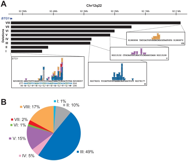
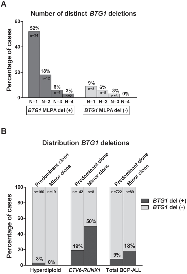
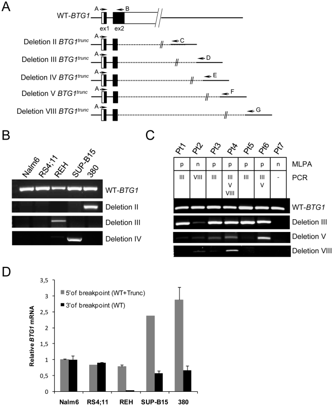
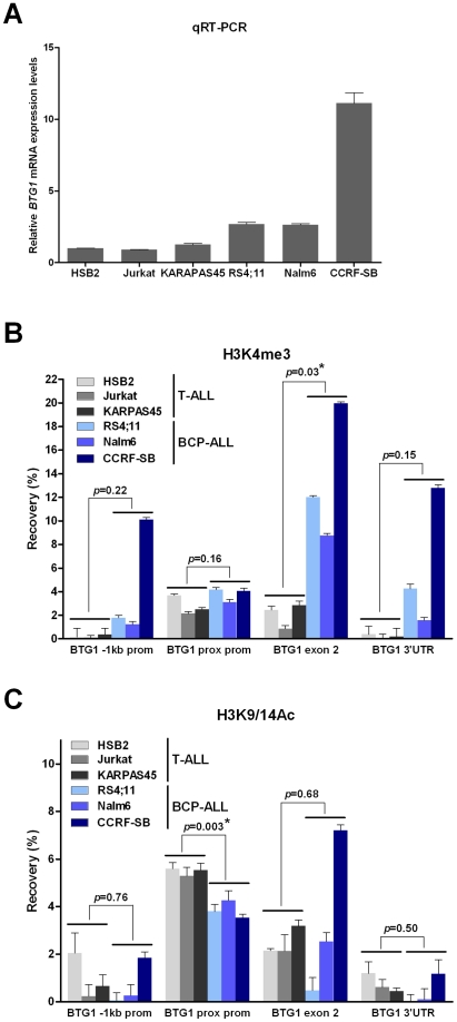
Recurrent submicroscopic deletions in genes affecting key cellular pathways are a hallmark of pediatric acute lymphoblastic leukemia (ALL). To gain more insight into the mechanism underlying these deletions, we have studied the occurrence and nature of abnormalities in one of these genes, the B-cell translocation gene 1 (BTG1), in a large cohort of pediatric ALL cases. BTG1 was found to be exclusively affected by genomic deletions, which were detected in 65 out of 722 B-cell precursor ALL (BCP-ALL) patient samples (9%), but not in 109 T-ALL cases. Eight different deletion sizes were identified, which all clustered at the telomeric site in a hotspot region within the second (and last) exon of the BTG1 gene, resulting in the expression of truncated BTG1 read-through transcripts. The presence of V(D)J recombination signal sequences at both sites of virtually all deletions strongly suggests illegitimate RAG1/RAG2-mediated recombination as the responsible mechanism. Moreover, high levels of histone H3 lysine 4 trimethylation (H3K4me3), which is known to tether the RAG enzyme complex to DNA, were found within the BTG1 gene body in BCP-ALL cells, but not T-ALL cells. BTG1 deletions were rarely found in hyperdiploid BCP-ALLs, but were predominant in other cytogenetic subgroups, including the ETV6-RUNX1 and BCR-ABL1 positive BCP-ALL subgroups. Through sensitive PCR-based screening, we identified multiple additional BTG1 deletions at the subclonal level in BCP-ALL, with equal cytogenetic distribution which, in some cases, grew out into the major clone at relapse. Taken together, our results indicate that BTG1 deletions may act as "drivers" of leukemogenesis in specific BCP-ALL subgroups, in which they can arise independently in multiple subclones at sites that are prone to aberrant RAG1/RAG2-mediated recombination events. These findings provide further evidence for a complex and multiclonal evolution of ALL.


Conflict of interest statement

The authors have declared that no competing interests exist.

Figures

Figure 1
Figure 1. BTG1 deletions cluster tightly and disrupt the BTG1 open reading frame.
(A) The BTG1 gene located on the antisense strand of chromosome 12q22 is exclusively disrupted by deletions (indicated by black bars). We identified 8 different deletions ranging from 101 to 557 kb in size. The deletion breakpoints cluster tightly within exon 2, the majority of which are located within a stretch of 10 base pairs. Colored triangles indicate the position of the breakpoints in different patients. Light blue represents the breakpoint of deletion I, grey deletion II, blue deletion III, pink deletion IV, purple deletion V, green deletion VI, red deletion VII and orange deletion VIII, respectively. The exact BTG1 deletion breakpoint sequences are listed in Table S2. Chromosomal location refers to human GRCh37/hg19 genome assembly. (B) In our cohort of 65 deletion positive BCP-ALL cases, as determined by MLPA, we detected eight distinct deletions with different frequencies. Deletion III was most prevalent (49% of the cases).
Figure 2
Figure 2. Multiple BTG1 deletion-positive clones are present in specific BCP-ALL subtypes.
(A) Recurrence of multiclonal BTG1 deletions. A sensitive PCR method was used to screen for eight different deletion breakpoints (deletion I–VIII) in BTG1 MLPA deletion positive (+) cases (n = 65), and to screen for the three most frequent deletion breakpoints (deletion III, V and VIII) in BTG1 MLPA deletion negative (−) cases (n = 89). (B) BTG1 deletion frequency in the two major cytogenetic subgroups of BCP-ALL (Hyperdiploid and ETV6-RUNX1). Presence of a BTG1 deletion in the predominant clone was determined by MLPA on the entire cohort of BCP-ALL cases (n = 722), and was compared to deletions detected as a minor clone in MLPA-negative cases (n = 89) by deletion-spanning PCR. Distributions are similar, being depleted from hyperdiploid cases and enriched in ETV6-RUNX1-positive cases as compared to the total group.
Figure 3
Figure 3. Expression of BTG1 truncated read-through transcripts in BCP-ALL cells with BTG1 deletions.
(A) Schematic representation of the wild-type human BTG1 gene, existing of two partly coding exons, and five different BTG1 transcripts due to BTG1 gene deletions. Exons are represented by black (coding) or white (non-coding) bars. Indicated are the RT-PCR primers that were used to detect expression of the wild-type BTG1 transcript (primers A and B), or one of the BTG1 truncated read-through transcripts for deletion II (primers A and C), deletion III (pimers A and D), deletion IV (primers A and E), deletion V (primers A and F), or deletion VIII (primers A and G). (B) RT-PCR analyses on total RNA isolated from the BCP-ALL cell lines Nalm6 and RS4;11 (BTG1 wild-type) and REH, SUP-B15 and 380, each with distinct monoallelic BTG1 deletions. (C) RT-PCR analyses on primary BCP-ALL samples in which a single BTG1 deletion (Pt1, Pt2, Pt3 and Pt5), multiple BTG1 deletions (Pt4 and Pt6) or no BTG1 deletions were detected with genomic PCR (Pt7). Type of deletions (III, V, or VIII) and outcome of MLPA (p: deletion-positive; n: deletion-negative) are indicated. BTG1 read-through transcripts were verified by sequencing (Table S5), except for Pt3-deletion V, which was an unrelated DNA sequence. (D) Quantitative real-time RT-PCR data representing relative expression levels of BTG1 measured 5′ (primers exon 1/2) and 3′ of the BTG1 breakpoint hotspot (primers exon 2). Expression levels were normalized to HPRT levels, and compared to the expression level in Nalm6, which was set to 1. The data shown represent the average of two independent cDNA reactions and triplicate qRT-PCR reactions.
Figure 4
Figure 4. Increased levels of H3K4me3 at the BTG1 locus in BCP-ALL versus T-ALL cell lines.
(A) Quantitative real-time RT-PCR data representing relative expression levels of BTG1 in T-ALL cell lines HSB2, Jurkat and KARPAS45, and BCP-ALL cell lines RS4;11, Nalm6 and CCRF-SB (HPRT normalized and related to HSB2 expression levels). Data shown are the average of two independent cDNA reactions and triplicate qRT-PCR reactions. (B and C) Percentage recovery after ChIP performed with H3K4me3 antibody (B) or H3K9/14Ac antibody (C) on T-ALL (HSB2, Jurkat, KARPAS45) and BCP-ALL (RS4;11, Nalm6 and CCRF-SB) cell lines. Real-time quantitative PCR was performed with primers specific for the region 1 kb upstream of the transcription start site (−1 kb prom), directly flanking the transcription start site (prox prom), the second exon near the breakpoint hotspot (exon 2) and towards the end of the 3′untranslated region (3′UTR) at the second (and last) exon of the human BTG1 gene. Values represent two independent ChIP experiments. Student's t-test was performed to assess differences between the average recovery of T-ALL versus BCP-ALL samples. Asterisk (*) indicates a p-value<0.05.

References

    1. Pui CH, Relling MV, Downing JR. Acute lymphoblastic leukemia. N Engl J Med. 2004;350:1535–1548. - PubMed
    1. Mullighan CG, Goorha S, Radtke I, Miller CB, Coustan-Smith E, et al. Genome-wide analysis of genetic alterations in acute lymphoblastic leukaemia. Nature. 2007;446:758–764. - PubMed
    1. Kuiper RP, Schoenmakers EF, Reijmersdal van SV, Hehir-Kwa JY, Geurts van Kessel A, et al. High-resolution genomic profiling of childhood ALL reveals novel recurrent genetic lesions affecting pathways involved in lymphocyte differentiation and cell cycle progression. Leukemia. 2007;21:1258–1266. - PubMed
    1. Tsuzuki S, Karnan S, Horibe K, Matsumoto K, Kato K, et al. Genetic abnormalities involved in t(12;21) TEL-AML1 acute lymphoblastic leukemia: analysis by means of array-based comparative genomic hybridization. Cancer Sci. 2007;98:698–706. - PMC - PubMed
    1. Kuiper RP, Waanders E, van der Velden VHJ, van Reijmersdal SV, Venkatachalam R, et al. IKZF1 deletions predict relapse in uniformly treated pediatric precursor B-ALL. Leukemia. 2010;24:1258–1264. - PubMed

Publication types

MeSH terms