. 2012;7(2):e27517.
doi: 10.1371/journal.pone.0027517. Epub 2012 Feb 16.

Decoding the distribution of glycan receptors for human-adapted influenza A viruses in ferret respiratory tract

Affiliations

Decoding the distribution of glycan receptors for human-adapted influenza A viruses in ferret respiratory tract

Akila Jayaraman et al. PLoS One. 2012.

Abstract

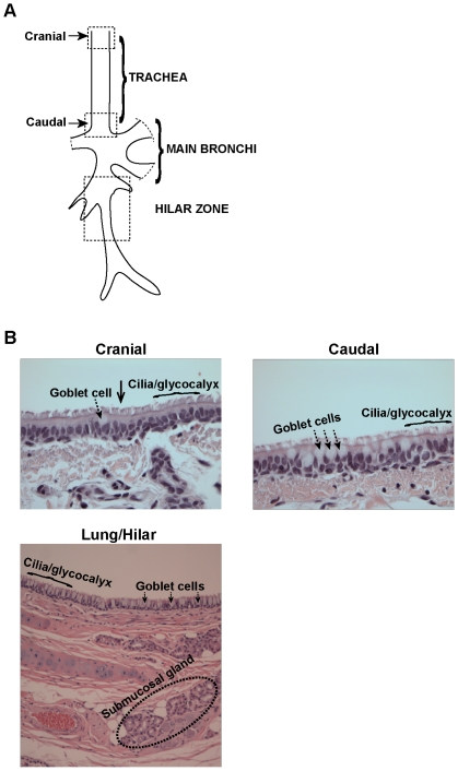
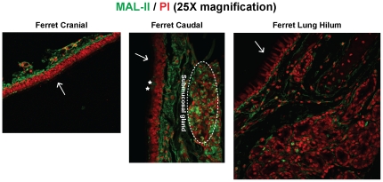
Ferrets are widely used as animal models for studying influenza A viral pathogenesis and transmissibility. Human-adapted influenza A viruses primarily target the upper respiratory tract in humans (infection of the lower respiratory tract is observed less frequently), while in ferrets, upon intranasal inoculation both upper and lower respiratory tract are targeted. Viral tropism is governed by distribution of complex sialylated glycan receptors in various cells/tissues of the host that are specifically recognized by influenza A virus hemagglutinin (HA), a glycoprotein on viral surface. It is generally known that upper respiratory tract of humans and ferrets predominantly express α2→6 sialylated glycan receptors. However much less is known about the fine structure of these glycan receptors and their distribution in different regions of the ferret respiratory tract. In this study, we characterize distribution of glycan receptors going beyond terminal sialic acid linkage in the cranial and caudal regions of the ferret trachea (upper respiratory tract) and lung hilar region (lower respiratory tract) by multiplexing use of various plant lectins and human-adapted HAs to stain these tissue sections. Our findings show that the sialylated glycan receptors recognized by human-adapted HAs are predominantly distributed in submucosal gland of lung hilar region as a part of O-linked glycans. Our study has implications in understanding influenza A viral pathogenesis in ferrets and also in employing ferrets as animal models for developing therapeutic strategies against influenza.


Conflict of interest statement

Competing Interests: The authors have declared that no competing interests exist.

Figures

Figure 1
Figure 1. Overview of ferret respiratory tract.
(A) Regions of ferret respiratory tract analyzed in this study. (B) Images of H&E stained ferret tracheal and lung hilar region. As seen from the images, the lung hilar region seems to have more goblet cells than the ferret trachea, a feature distinct from human respiratory tract.
Figure 2
Figure 2. α2–3 linked glycan distribution in ferret respiratory tract.
MAL-II lectin (green) was used to stain ferret cranial, caudal and lung hilar regions. As seen from the images, MAL-II stained the submucosal glands, the underlying mucosa and some goblet cells (marked as *) in the caudal region. There was no staining of the lung hilar region indicating an absence of α2–3 glycans. The nuclei were stained with PI (red). The apical surface is marked with a white arrow.
Figure 3
Figure 3. O linked α2–6 glycan distribution in ferret respiratory tract.
A, Staining of ferret cranial, caudal and lung hilar regions with FITC-labeled SNA-I (green). As seen from the images, SNA-I did not stain any of the goblet cells in the ferret trachea (cranial and caudal) although some of the goblet cells (marked as *) in the lung hilar region were stained by SNA-I. Submucosal glands and glycocalyx in both ferret trachea and lung hilar regions showed significant staining with SNA-I. B, Staining of ferret cranial, caudal and lung hilar regions with FITC-labeled Jacalin (green); a plant lectin having specificity to Tn antigen. Jacalin stained the goblet cells (marked as *), and the submucosal glands in all the three regions of ferret respiratory tract similar to human trachea. C, Co-staining of ferret respiratory tissue sections with FITC-labeled Jacalin (green) and SNA-I tagged to a 546 nm fluorophore (red). Co-staining is seen as yellow color due to the overlap of green (Jacalin) and red (SNA) fluorophores. Submucosal glands in both the trachea and lung hilar region showed significant co-staining with Jacalin/SNA-I. There was some co-staining of goblet cells in the ferret lung hilar region (data not shown) and in ferret trachea cranial region (marked as *). In case of staining with single lectin, the nuclei were visualized by staining with PI (red). The apical surface is marked with a white arrow.
Figure 4
Figure 4. Glycan receptor distribution for human-adapted influenza A virus HA.
Recombinant SC18 (H1) HA and Alb58 (H2) HA expressed in insect cells were used to stain ferret cranial, caudal and lung hilar regions at a concentration of 20 µg/ml. A, SC18 HA stained only the submucosal glands in all the three regions of ferret respiratory tract. This is in contrast to human trachea where goblet cells (marked as *) were stained by SC18 HA. This restricted binding pattern of SC18 HA can be attributed to its stringent binding specificity to long α2–6 linked (6′SLNLN) glycans. B, Alb58 HA stained the submucosal glands, the underlying mucosa and some goblet cells (in the caudal region) similar to SNA-I. This staining pattern is similar to that in human trachea wherein all the goblet cells (marked as *), submucosal glands and the glycocalyx are stained with Alb58HA. The significant goblet cell staining of Alb58 HA of human trachea as compared to ferret respiratory tract is in accordance with predominant expression of O-linked α2–6 sialic acid in human tracheal goblet cells as compared to that in ferret respiratory tract ( Figure 3 ). The nuclei were stained with PI (red). The apical surface is marked with a white arrow.
Figure 5
Figure 5. Co-staining of recombinant Alb58 (H2) HA with Jacalin.
Ferret cranial, caudal and lung hilar regions were co-stained with 10 µg/ml of Jacalin (FITC labeled) and 20 µg/ml of recombinant Alb58 HA. The co-staining is indicated by a yellow staining pattern. As seen from the images, co-staining was predominantly seen in the submucosal glands of ferret trachea and lung hilar region. Some co-staining was also seen in the goblet cells of trachea (marked as *) and lung hilar region. This staining pattern is in contrast to human trachea wherein co-staining is predominantly seen in goblet cells (marked as *). The apical surface is marked with a white arrow.
Figure 6
Figure 6. Glycan receptor distribution in ferret respiratory tract.
Shown in the figure is the glycan receptor distribution in ferret in ferret trachea (A) and lung hilar region (B) based on lectin staining patterns. The glycan receptor motifs are shown in cartoon representation (see Table S1 for cartoon key). Note that predominant α2–6 glycan receptors recognized by human influenza A viruses are found in the submucosal glands of trachea and lung hilum. Further, unlike human respiratory tract, there is minimal to no α2–3 sialylated glycan receptor expression in the lung hilar region of ferret. Except for differences in the number of goblet cells in ferret cranial and caudal region, there were no significant differences in the glycan receptor expression. The apical surface is marked with a black arrow.

Similar articles

Cited by

References

    1. van Riel D, Munster VJ, de Wit E, Rimmelzwaan GF, Fouchier RA, et al. Human and Avian Influenza Viruses Target Different Cells in the Lower Respiratory Tract of Humans and Other Mammals. Am J Pathol. 2007;171:1215–1223. - PMC - PubMed
    1. van Riel D, den Bakker MA, Leijten LM, Chutinimitkul S, Munster VJ, et al. Seasonal and pandemic human influenza viruses attach better to human upper respiratory tract epithelium than avian influenza viruses. Am J Pathol. 2010;176:1614–1618. - PMC - PubMed
    1. Belser JA, Katz JM, Tumpey TM. The ferret as a model organism to study influenza A virus infection. Dis Model Mech. 2011;4:575–579. - PMC - PubMed
    1. Maher JA, DeStefano J. The ferret: an animal model to study influenza virus. Lab Anim (NY) 2004;33:50–53. - PubMed
    1. Tumpey TM, Maines TR, Van Hoeven N, Glaser L, Solorzano A, et al. A two-amino acid change in the hemagglutinin of the 1918 influenza virus abolishes transmission. Science. 2007;315:655–659. - PubMed

Publication types

Substances