. 2012;7(2):e29727.
doi: 10.1371/journal.pone.0029727. Epub 2012 Feb 16.

Rheotaxis in larval zebrafish is mediated by lateral line mechanosensory hair cells

Affiliations

Rheotaxis in larval zebrafish is mediated by lateral line mechanosensory hair cells

Arminda Suli et al. PLoS One. 2012.

Abstract

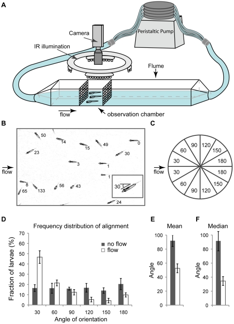
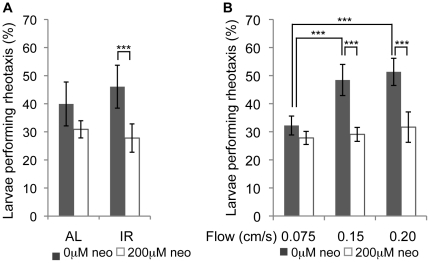
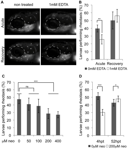
The lateral line sensory system, found in fish and amphibians, is used in prey detection, predator avoidance and schooling behavior. This system includes cell clusters, called superficial neuromasts, located on the surface of head and trunk of developing larvae. Mechanosensory hair cells in the center of each neuromast respond to disturbances in the water and convey information to the brain via the lateral line ganglia. The convenient location of mechanosensory hair cells on the body surface has made the lateral line a valuable system in which to study hair cell damage and regeneration. One way to measure hair cell survival and recovery is to assay behaviors that depend on their function. We built a system in which orientation against constant water flow, positive rheotaxis, can be quantitatively assessed. We found that zebrafish larvae perform positive rheotaxis and that, similar to adult fish, larvae use both visual and lateral line input to perform this behavior. Disruption or damage of hair cells in the absence of vision leads to a marked decrease in rheotaxis that recovers upon hair cell repair or regeneration.


Conflict of interest statement

Competing Interests: The authors have declared that no competing interests exist.

Figures

Figure 1
Figure 1. Zebrafish larvae can rheotax in flow conditions.
A) We built a clear plastic, rectangular flume in which to test rheotaxis. 20–30 larvae are placed in observation chamber and their alignment is recorded. B) Using ImageJ (NIH) macros, images are cropped, ellipses are fitted around each larva, and their alignment angle against current is calculated. Larvae display a preference for aligning against current in flow conditions (0.14 cm/s) as shown by frequency distribution (D), mean (E) and median (F) of alignment. C) Larvae show no left-right preference; therefore, their alignment is calculated in a 0–180° scale (C). Experiment performed in IR.
Figure 2
Figure 2. Zebrafish larvae use the lateral line system to align in flow.
Alignment of larvae at 30° is shown. A) Removing visual cues and lateral line input leads to reduced rheotaxis (n = 5) (flow = 0.16 cm/s). AL: ambient light illumination; sides of flume covered by paper with vertical stripes; IR: infrared illumination. B) Untreated larvae align better with increased flow rates, while alignment of lateral-line-impaired larvae does not change significantly at different flow rates (n = 5). Statistics: ANOVA with Tukey post-test. Error bars show standard deviation. *** p<0.001.
Figure 3
Figure 3. Rheotaxis depends on level of hair cell impairment or damage and is restored during regeneration.
A) Mechanotransduction, as shown by FM1–43 dye uptake, is blocked when tip-links are broken at ∼30 min after EDTA treatment (acute) and is restored again at 4–5 hours post treatment (recovery) (flow = 0.16 cm/s). B) Rheotaxis is impaired acutely after EDTA treatment and restored to control levels after recovery (n = 5). C) Ablating the lateral line input by different neomycin doses leads to a dose dependent rheotaxis impairment (n = 6) (flow = 0.14 cm/s). D) Rheotaxis is restored during mechanosensory hair cell regeneration (n = 5) (flow = 0.16 cm/s). Statistics: C) Statistics: ANOVA with Tukey post-tests. B,D) Paired t-test. Error bars show standard deviation. * p = 0.01–0.05, ** p = 0.01–0.001, *** p<0.001.

Similar articles

Cited by

References

    1. Ghysen A, Dambly-Chaudiere C. The lateral line microcosmos. Genes Dev. 2007;21:2118–2130. - PubMed
    1. Metcalfe WK, Kimmel CB, Schabtach E. Anatomy of the posterior lateral line system in young larvae of the zebrafish. J Comp Neurol. 1985;233:377–389. - PubMed
    1. Balak KJ, Corwin JT, Jones JE. Regenerated hair cells can originate from supporting cell progeny: evidence from phototoxicity and laser ablation experiments in the lateral line system. J Neurosci. 1990;10:2502–2512. - PMC - PubMed
    1. Partridge BL, Pitcher TL. The sensory basis of fish schools; relative roles of lateral line and vision. J Comp Physiol. 1980;135:315–325.
    1. Hoekstra D, Janssen J. Non-visual feeding behaviour of the mottled sculpin, Cottus bairdi in Lake Michigan. Env Biol Fish. 1985;12:111–117.

Publication types