. 2012;7(2):e30512.
doi: 10.1371/journal.pone.0030512. Epub 2012 Feb 16.

Improved control of tuberculosis and activation of macrophages in mice lacking protein kinase R

Affiliations

Improved control of tuberculosis and activation of macrophages in mice lacking protein kinase R

Kangyun Wu et al. PLoS One. 2012.

Erratum in

Abstract

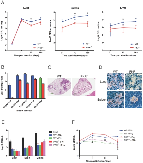
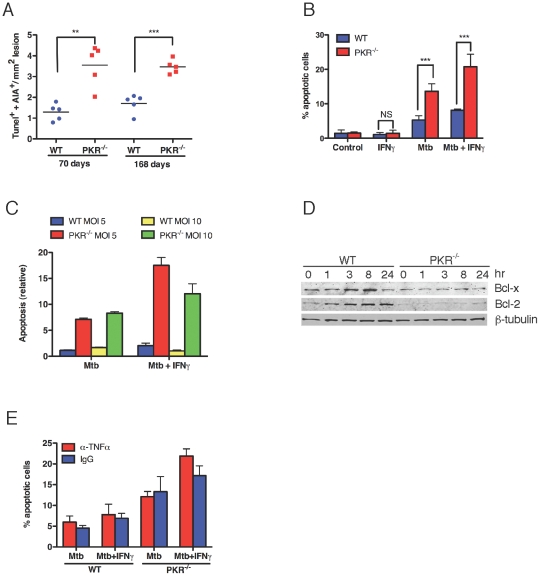
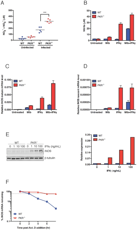
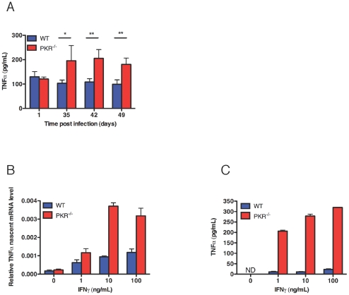
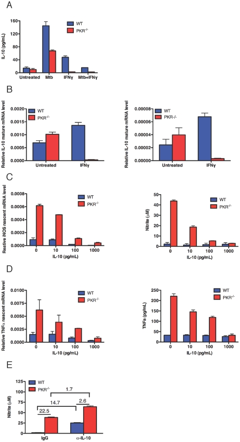
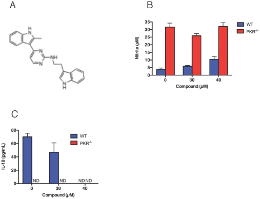
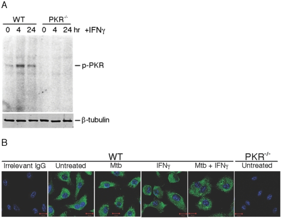
Host factors that microbial pathogens exploit for their propagation are potential targets for therapeuic countermeasures. No host enzyme has been identified whose genetic absence benefits the intact mammalian host in vivo during infection with Mycobacterium tuberculosis (Mtb), the leading cause of death from bacterial infection. Here, we report that the dsRNA-dependent protein kinase (PKR) is such an enzyme. PKR-deficient mice contained fewer viable Mtb and showed less pulmonary pathology than wild type mice. We identified two potential mechanisms for the protective effect of PKR deficiency: increased apoptosis of macrophages in response to Mtb and enhanced activation of macrophages in response to IFN-gamma. The restraining effect of PKR on macrophage activation was explained by its mediation of a previously unrecognized ability of IFN-gamma to induce low levels of the macrophage deactivating factor interleukin 10 (IL10). These observations suggest that PKR inhibitors may prove useful as an adjunctive treatment for tuberculosis.


Conflict of interest statement

Competing Interests: The authors have declared that no competing interests exist.

Figures

Figure 1
Figure 1. Course of Mtb infection in PKR-deficient and wild type mice.
(A) Time course of burden of colony-forming units (CFU) in lung, liver and spleen over 24 weeks following infection by inhalation of an average of 30 CFU by wild type mice and 37 CFU by PKR-deficient mice. Results are means ± SD for 5 mice per time point. Asterisks mark time points at which the differences had p values≤0.01 by Student's t test. (B) Mtb burdens (mean CFU ± SEM for 4–6 mice per strain) in lungs of mice from 5 independent experiments at the latest time point post-infection evaluated in each experiment. Red bars: PKR−/− mice. Blue bars: C57LB/6 wild type controls. Green bar: 129S1/SvImJ wild type controls. The initial bacterial burdens were comparable between mouse strains within each experiment as assessed by the following CFU counts 24 hours post infection (pairs are means for wild type mice followed by PKR-deficient mice in individual experiments in the order depicted in the figure): 30, 37; 15, 20; 24, 22; 488, 468; 40, 45. The unusually low CFU seen in the first experiment are depicted with reference to an expanded Y-axis. (C and D) Histopathology (C) and acid-fast staining of Mtb (D) in lungs from Mtb-infected wild type and PKR−/− mice at day 168 from a representative experiment. (E and F) Comparable uptake and control of Mtb by PKR-deficient and wild type macrophages in vitro. (E) Uptake of Mtb 4 hours after addition at the indicated MOI, with and without exposure of macrophages to IFN-gamma (10 ng/mL) overnight in advance of infection. (F) Intracellular growth of Mtb (MOI = 3) with and without exposure of macrophages to IFN-gamma (10 ng/mL) overnight in advance of infection. With the other MOIs tested (1 and 10), there was likewise no significant difference in the numbers of CFU in PKR-deficient and wild type macrophages over the 3 days studied.
Figure 2
Figure 2. Enhanced apoptosis of the PKR-deficient macrophages.
(A) Apoptosis of macrophages in vivo. After 70 or 168 days of infection, sections of lungs from 5 WT and 5 PKR−/− mice were stained by TUNEL and with an anti-macrophage antibody, AIA . TUNEL-positive macrophages were counted in 4 microscopic fields per lung in areas of comparable histopathology such that approximately 20–40 TUNEL-positive cells were evaluated per mouse. **, p<0.005, ***; p<0.0005 (unpaired t test). (B) Apoptosis of macrophages in vitro assessed by TUNEL. Macrophages (5×10⁴) from wild type and PKR−/− mice were incubated with or without IFNγ (10 ng/mL) overnight and then infected with Mtb at MOI 10. After 24 h, apoptosis was assayed by counting the proportion of TUNEL-positive macrophages in 4 microscopic fields per well. Control, no Mtb. ***, p≤0.0005 (unpaired t test). NS, not significant. (C) Apoptosis of macrophages in vitro assessed by ELISA for cytoplasmic histone-associated mono- and oligo-nucleosomes. Macrophages were infected as in (B). The relative extent of apoptosis is presented as a ratio of absorbance values from infected macrophages to those from uninfected macrophages. In (B) and (C), results are means ± SD for 3 replicates in one experiment representative of 2. (D) Western blot for apoptosis-inhibitory proteins at the indicated times after infection of macrophages with Mtb at MOI = 1. Immunoblot for β-tubulin served as a loading control. (E) Effect of anti-TNF-alpha on apoptosis of primary macrophages in response to Mtb infection. Macrophages from WT and PKR−/− mice were incubated with or without IFN-gamma (10 ng/mL) overnight and then treated with anti–TNF-alpha IgG (10 µg/mL) or isotype-matched irrelevant IgG just before the infection with Mtb at MOI 10 for 24 hr. Apoptosis was assayed by staining with TUNEL and quantified as the percentage of TUNEL positive cells among DAPI positive cells. Data are means ± SD for 4 fields of cells for each condition.
Figure 3
Figure 3. Enhanced expression of iNOS in PKR-deficient mice and macrophages in response to Mtb and IFN-gamma.
(A) Nitrite plus nitrate in sera from Mtb-infected wild type and PKR-deficient mice. Nitrite and nitrate in serum from Mtb-infected mice at day 168 were measured by the Griess reaction after nitrate was reduced to nitrite as described . Means ± SD of 4–5 mice in one experiment representative of two. ***, p<0.0005 (unpaired t test). (B) Secretion of nitrite by macropahges in vitro. Macrophages (5×10⁵) were treated with Mtb (MOI 3), IFN-gamma (10 ng/mL), or pre-treated with IFN-gamma (10 ng/mL) overnight followed by infection with Mtb (MOI 3) for 24 h. Nitrite in the supernatant was determined by the Griess reaction. Means ± SD of triplicates. (C and D) Mature and nascent iNOS transcripts. Macrophages were incubated as in (B) for 3 h. qRT-PCR signals for iNOS were normalized to GAPDH. Means ± SD of triplicates. In (B), (C) and (D), results are from one of two similar experiments. Results were similar in additional experiments at MOI's of 0.3, 1 and 3 and IFN-gamma concentrations of 1, 10 and 100 ng/mL. (E) Western blot (left) and its densitometric assessment (right) for iNOS in macrophages incubated with the indicated concentrations of IFN-gamma for 24 h. Immunoblot for beta-tubulin served as a loading control. (F) Impact of PKR deficiency on stability of iNOS mRNA. Macrophages were treated with IFN-gamma (10 ng/mL) followed 3 h later by actinomycin D (10 micrograms/mL). RNA was extracted at the indicated times for RT-PCR and normalized to GAPDH.
Figure 4
Figure 4. Enhanced expression of TNF-alpha in PKR-deficient mice and macrophages.
(A) Levels of TNF-alpha in lung homogenates. TNF-alpha in lung homogenates from Mtb-infected mice at indicated time was measured by ELISA. Means ± SD for 4 mice per strain from one experiment representative of four. *, p<0.05, **, p<0.005, (unpaired t test). (B) Nascent TNF-alpha transcripts. Macrophages (5×10⁵) were incubated with the indicated concentrations of IFN-gamma for 3 h. qRT-PCR signals for TNF-alpha were normalized to GAPDH. Means ± SD of triplicates. (C) Secretion of TNF-alpha. Macrophages (5×10⁵) were incubated with the indicated concentrations of IFN-gamma for 48 h. Supernatant was collected for TNF-alpha production by ELISA. Means ± SD of triplicates. In (B) and (C), results are from one of two similar experiments.
Figure 5
Figure 5. Induction of IL-10 by Mtb and IFN-gamma and its impact on iNOS and TNF-alpha.
(A) Secretion of IL-10. Macrophages (5×10⁵) were treated with Mtb (MOI 3), IFN-gamma (10 ng/mL), or pre-treated with IFN-gamma (10 ng/mL) overnight followed by infection with Mtb (MOI 3) for 24 h. Supernatant was collected for IL-10 production by ELISA. Results were similar in additional experiments with MOIs of 0.3, 1 and 3 and IFN-gamma concentrations of 1, 10 and 100 ng/mL. (B) Induction of mature and nascent IL-10 transcripts. Macrophages (5×10⁵) were treated with IFN-gamma (10 ng/mL) for 3 h. qRT-PCR signals for IL-10 were normalized to GAPDH. Results were similar with IFN-gamma concentrations of 1, 10 and 100 ng/mL. (C) Effect of IL-10 on nascent iNOS transcripts and secretion of nitrite. Left panel, macrophages (5×10⁵) were treated with IL-10 for 3 h after the addition of IFN-gamma 0 ng/mL. qRT-PCR signals for iNOS were normalized to GAPDH. Right panel, macrophages (5×10⁵) were treated with IL-10 for 48 h after the addition of IFN-gamma 0 ng/mL. Nitrite in the supernatant was measured by the Griess reaction. (D) Effect of IL10 on nascent TNF-alpha transcripts and TNF-alpha. Left panel, macrophages (5×10⁵) were treated with IL-10 for 3 h after the addition of IFN-gamma 0 ng/mL. qRT-PCR signals for TNF-alpha were normalized to GAPDH. Right panel, macrophages (5×10⁵) were treated with IL-10 for 48 h after the addition of IFN-gamma 0 ng/mL. TNF-alpha in the supernatant was measured by ELISA. (E) Effect of neutralizing anti-IL-10 antibody (10 µg/mL) on release of nitrite 48 h after addition of IFN-gamma (10 ng/mL). Results in (A) to (F) are means ± SD from individual experiments each with 3 replicates that are representative of at least 2 independent experiments.
Figure 6
Figure 6. A PKR inhibitor partially phenocopies the effect of PKR deficiency on release of nitrite and IL10 in response to IFN-gamma.
(A) Structure of the PKR inhibitor N-(2(1H-indol-3-yl)ethyl)-4-(2-methyl-1H-indol-3-yl)pyrimin-2-amine. (B) Impact of the inhibitor on nitrite release 48 h after addition of IFN-gamma (10 ng/mL) to wild type macrophages. (C) Impact on IL10 secretion 48 h after addition of IFN-gamma (10 ng/mL) to wild type macrophages. (B) and (C) are means ± SD for 3 replicates in one of 3 similar experiments.
Figure 7
Figure 7. Activation of PKR by IFN-gamma.
(A) Autophosphorylation of PKR in macrophages at the indicated times after addition of IFN-gamma (10 ng/mL). A duplicate gel western blotted with antibody to beta-tubulin served as a loading control. One of four similar experiments. (B) Immunofluorescent localization of PKR in wild type macrophages with and without Mtb infection and/or IFN-gamma treatment. Macrophages from wild type and PKR−/− mice were incubated with or without IFN-gamma (10 ng/mL) at 37°C overnight and then infected with Mtb at MOI 10 for 24 h. Macrophages were fixed with 4% p-formaldehyde for 2 h and immunofluorescent staining for PKR (green) and DNA (DAPI; blue) was conducted. Images were recorded by confocal microscopy.

Similar articles

Cited by

References

    1. Schwegmann A, Brombacher F. Host-directed drug targeting of factors hijacked by pathogens. Sci Signal. 2008;1:re8. - PubMed
    1. Chang AC, Zsak L, Feng Y, Mosseri R, Lu Q, et al. Phenotype-based identification of host genes required for replication of African swine fever virus. J Virol. 2006;80:8705–8717. - PMC - PubMed
    1. Cohen SN. Microbial drug resistance: An old problem in need of new solutions. In: Relman DA, Hamburg MA, Choffnes EE, Mack A, editors. Microbial Evolution and Co-Adaptation:A Tribute to the Life and Scientific Legacies of Joshua Lederberg. National Academies Press; 2009. pp. 173–180. 2009; Washington, DC. - PubMed
    1. Philips JA, Rubin EJ, Perrimon N. Drosophila RNAi screen reveals CD36 family member required for mycobacterial infection. Science. 2005;309:1251–1253. - PubMed
    1. Kuijl C, Savage ND, Marsman M, Tuin AW, Janssen L, et al. Intracellular bacterial growth is controlled by a kinase network around PKB/AKT1. Nature. 2007;450:725–730. - PubMed

Publication types

MeSH terms