. 2012;7(2):e31218.
doi: 10.1371/journal.pone.0031218. Epub 2012 Feb 16.

The functions of myosin II and myosin V homologs in tip growth and septation in Aspergillus nidulans

Affiliations

The functions of myosin II and myosin V homologs in tip growth and septation in Aspergillus nidulans

Naimeh Taheri-Talesh et al. PLoS One. 2012.

Abstract

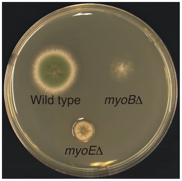
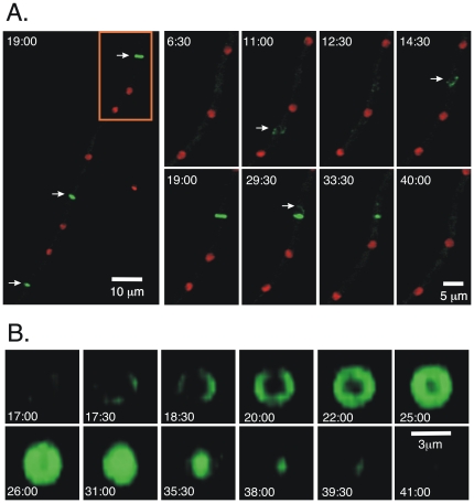
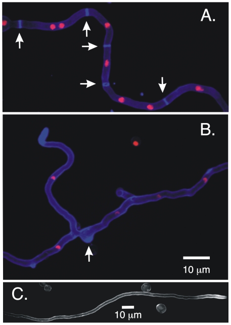
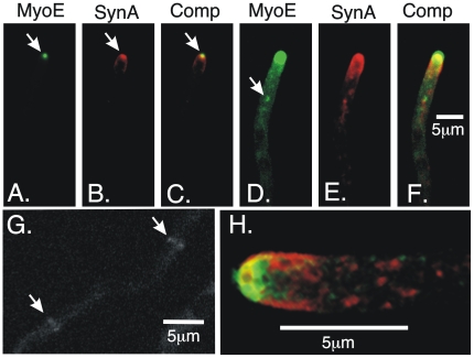
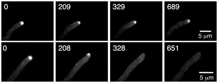
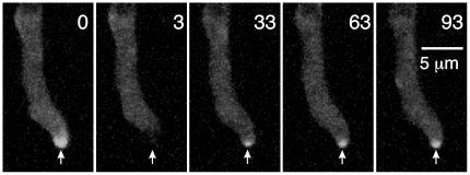
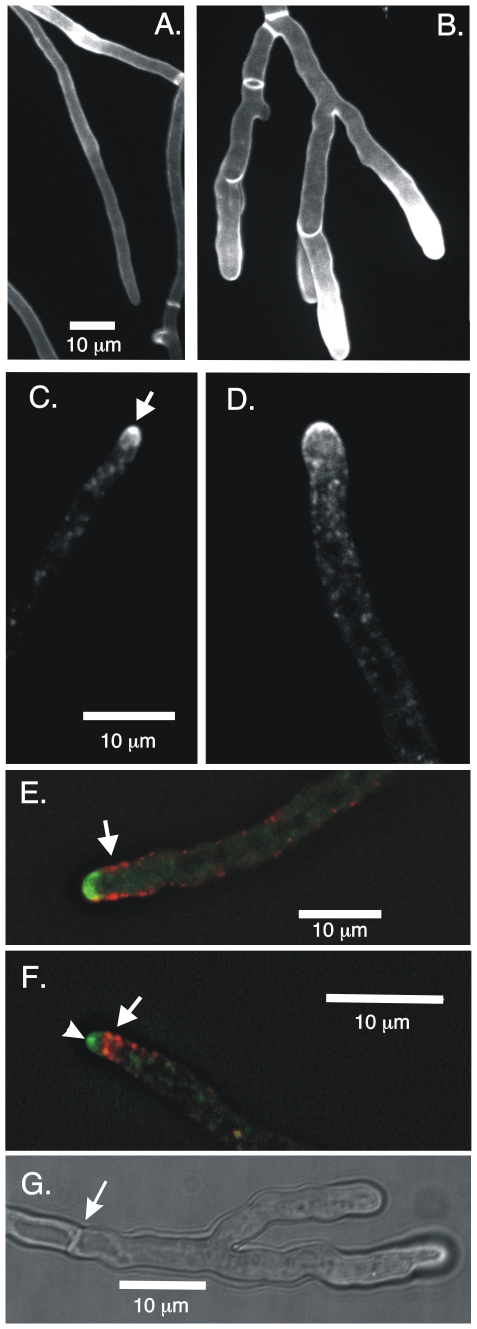
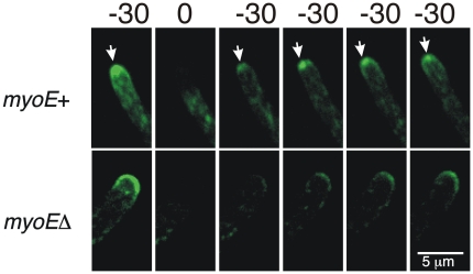
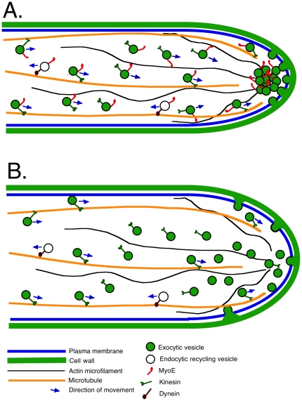
Because of the industrial and medical importance of members of the fungal genus Aspergillus, there is considerable interest in the functions of cytoskeletal components in growth and secretion in these organisms. We have analyzed the genome of Aspergillus nidulans and found that there are two previously unstudied myosin genes, a myosin II homolog, myoB (product = MyoB) and a myosin V homolog, myoE (product = MyoE). Deletions of either cause significant growth defects. MyoB localizes in strings that coalesce into contractile rings at forming septa. It is critical for septation and normal deposition of chitin but not for hyphal extension. MyoE localizes to the Spitzenkörper and to moving puncta in the cytoplasm. Time-lapse imaging of SynA, a v-SNARE, reveals that in myoE deletion strains vesicles no longer localize to the Spitzenkörper. Tip morphology is slightly abnormal and branching occurs more frequently than in controls. Tip extension is slower than in controls, but because hyphal diameter is greater, growth (increase in volume/time) is only slightly reduced. Concentration of vesicles into the Spitzenkörper before incorporation into the plasma membrane is, thus, not required for hyphal growth but facilitates faster tip extension and a more normal hyphal shape.


Conflict of interest statement

Competing Interests: The authors have declared that no competing interests exist.

Figures

Figure 1
Figure 1. Growth phenotype of myosin deletants.
Incubation was for three days at 37°C on YAG medium supplemented with riboflavin. While both myoB and myoE deletants are viable, the myoB deletant colony is thin and wispy. Microscopic examination revealed that individual hyphae extend beyond the apparent edge of the colony. The myoE deletant is compact, exhibiting slower radial growth than the control strain.
Figure 2
Figure 2. MyoB-GFP localization during septum formation.
Images are projections from a time-lapse data set taken with strain LO2390. Times are in min and sec after the start of imaging. In A, the panel at the left is a low magnification image from the 19:00 min time point showing MyoB-GFP localization at three forming septa (arrows). Higher magnification time-lapse images of the region in the rectangle are shown to the right. The time after the beginning of time-lapse acquisition (in min) is shown at the upper left of each panel. In this set of images, strings of MyoB-GFP begin to coalesce at the 11 min time point (arrow) but then disperse and coalesce at a different place (arrow at 14:30). A septum then forms and begins to contract. Strings of MyoB-GFP can be seen leaving the septum (arrow at 29:30). Panel B is from the same time-lapse data set as A, but a single septum is shown and it is rotated 90° (using Volocity software) such that we have an end-on view of septum formation. MyoB-GFP assembles into a ring, with no evidence of it being spun out from a single spot. The ring then fills and contracts before disappearing. The time (in min) after the beginning of acquisition is shown at the bottom of each panel.
Figure 3
Figure 3. Deletion of myoB inhibits septum formation.
All panels are images of living cells. In A and B, nuclei are shown with histone H1-mRFP and chitin is stained with calcofluor (10 µg/ml). A. a myoB + strain (LO1516). Multiple septa are visible (arrows). B. a myoBΔ hypha. The myoB gene was deleted in LO1516 and nuclei carrying the deletion were maintained in a heterokaryon. No septa are present but there are thickened regions containing chitin (e.g. arrow) and chitin is highly concentrated near the hyphal tip. C. Shows a hyhal tip region in a myoBΔ strain stained with calcofluor but nuclei are not imaged. Note the absence of septa and side branches. The circular objects are ungerminated conidia resulting from the heterokaryon rescue technique.
Figure 4
Figure 4. MyoE localization.
A–F are images of the same field and are maximum intensity projections of a Z-series stack. A–C show the co-localization of MyoE-GFP and mCherry-SynA at the Spitzenkörper (arrows). SynA localizes to the Spitzenkörper and to the plasma membrane near the apex (B). In D–F, the thresholds are chosen to reveal the punctate staining in the hypha while overexposing the MyoE-GFP and mCherry-SynA at the hyphal tip. MyoE-GFP localizes to numerous small puncta and some larger structures that may be endosomes (e.g. arrow). G. Faint localization of MyoE-GFP at forming septa (arrows). H. A three-dimensional projection of a hyphal tip showing MyoE-GFP and mCherry-SynA. Although MyoE and SynA co-localize at the Spitzenkörper, many puncta behind the tip show GFP fluorescence or mCherry fluorescence, but it was not clear that there was any obligate co-localization.
Figure 5
Figure 5. Cytochalasin A causes MyoE-GFP to disperse from the Spitzenkörper.
Images are maximum intensity projections of Z-series stacks. Time (in sec) after the addition of DMSO (top row) or an equivalent volume of cytochalasin A dissolved in DMSO to give a final concentration of 1 µg/ml (bottom row). MyoE-GFP continuously localizes to the Spitzenkörper in the solvent control (top row) but disperses in less than 328 sec after the addition of cytochalasin A.
Figure 6
Figure 6. Movement of MyoE-GFP to the Spitzenkörper in the absence of microtubules.
Microtubules have been depolymerized with 2.4 µg/ml benomyl time at the upper right in each panel is in seconds. At t = 0, MyoE-GPF is visible at the Spitzenkörper (arrow). Three seconds later after FRAP the MyoE-GFP in the Spitzenkörper is bleached. In spite of the absence of microtubules, MyoE-GFP has moved to the Spitzenkörper 30 sec after FRAP (arrow, t = 33) and it increases in intensity at the Spitzenkörper over the next minute (arrows).
Figure 7
Figure 7. Deletion of myoE alters hyphal morphology and SynA distribution but not the localization of endocytic patches.
Panel A shows a myoE + strain and panel B shows a myoEΔ strain. Both are stained with 10 µg/ml calcofluor. Hyphae in the myoEΔ strain are thicker, vary more in thickness and exhibit more branching near the tip. The amount of chitin staining at the hyphal tip varied from hypha to hypha in wild-type strains as well as myoB and myoE deletion strains. The difference in staining between A and B is not specific to myoEΔ. Panel C shows GFP-SynA in a myoE + strain. SynA is concentrated into the Spitzenkörper at the hyphal tip (arrow) and is also present at the membrane near the tip. Panel D shows GFP-SynA in a myoEΔ strain. SynA is present at the membrane and in puncta in the cytoplasm but is not obviously organized into a Spitzenkörper. Panel E shows the localization of AbpA-mRFP and GFP-SynA in a myoEΔ strain. The image is a single focal plane from a deconvolved Z-series stack. AbpA-containing endocytic patches (arrow) localize to the cortex behind the growing tip and in three dimensions form a collar behind the growing tip. Panel F shows a control myoE + strain (LO1548) also expressing GFP-SynA and AbpA-mRFP. The image is a single focal plane from a deconvolved Z-series stack. The ApbA-containing patches (arrow) appear to be organized into a tighter array and the Spitzenkörper is visible (arrowhead). Note that myoE + hyphae are more consistent in diameter along their length than myoEΔ hyphae (compare A and B) and that the apices in myoEΔ hyphae appear rounder than in myoE + hyphae. A and B are the same magnification as are C and D. Panel G shows branching ahead of the first septum (septum designated with an arrow).
Figure 8
Figure 8. Fluorescence recovery after photobleaching (FRAP) of GFP-SynA in myoE+ and myoEΔ strains.
The tips of the two strains were photobleached at T = 0 (sec). In the myoE+ strain recovery is rapid. GFP-SynA appears at the tip within 30 sec of photobleaching and quickly localizes to the Spitzenkörper (arrows). This indicates that vesicles with SynA move rapidly to the tip and move through the Spitzenkörper before fusing with the plasma membrane. In the myoEΔ strain, the GFP-SynA is also visible at the tip at 30 sec after bleaching. MYOE, thus is not required for movement of GFP-SynA-containing vesicles to the tip. The GFP-SynA does not go through the Spitzenkörper, moreover, but fuses with the plasma membrane in a broad region of the tip.
Figure 9
Figure 9. A simplified model for MyoE function at the hyphal tip.
A. A myoE + cell. Exocytic vesicles move along microtubules powered by kinesin molecules. (It is likely that several kinesins can carry out this function.) There is a large zone of overlap between microtubules and actin microfilaments. When exocytic vesicles become detached from microtubules, as will generally be the case because of the limited processivity of kinesins, MyoE, on the vesicles will move the vesicles along actin microfilaments, collecting them at the Spitzenkörper. The vesicles then fuse in a fairly small area to the plasma membrane releasing their contents and resulting in hyphal growth. MyoE, vesicle components and, probably, many more proteins are moved in retrograde direction by dynein where they will be reused. B. A myoEΔ cell. In the absence of MyoE, exocytic vesicles are not focused into the Spitzenkörper but they are still moved into the hyphal apex area where they fuse with the plasma membrane over a wider area, resulting in hyphae with a greater diameter and lower extension rate. For simplicity, much of the endocytic machinery including endosomes and actin patches has been left out of this model. For a more detailed model of the endocytic machinery please see reference 10.

Similar articles

Cited by

References

    1. Harris SD, Morrell JL, Hamer JE. Identification and characterization of Aspergillus nidulans mutants defective in cytokinesis. Genetics. 1994;136:517–532. - PMC - PubMed
    1. McGoldrick CA, Gruver C, May GS. myoA of Aspergillus nidulans encodes an essential myosin I required for secretion and polarized growth. J Cell Biol. 1995;128:577–587. - PMC - PubMed
    1. Wolkow TD, Harris SD, Hamer JE. Cytokinesis in Aspergillus nidulans is controlled by cell size, nuclear positioning and mitosis. J Cell Sci. 1996;109:2179–2188. - PubMed
    1. Yamashita RA, May GS. Constitutive activation of endocytosis by mutation of myoA, the myosin I gene of Aspergillus nidulans. J Biol Chem. 1998;273:14644–14648. - PubMed
    1. Yamashita RA, Osherov N, May GS. Localization of wild type and mutant class I myosin proteins in Aspergillus nidulans using GFP-fusion proteins. Cell Motil Cytoskeleton. 2000;45:163–172. - PubMed

Publication types

MeSH terms

LinkOut - more resources