

Glucose enhances leptin signaling through modulation of AMPK activity
- PMID: 22359610
- PMCID: PMC3281098
- DOI: 10.1371/journal.pone.0031636
Glucose enhances leptin signaling through modulation of AMPK activity
Abstract
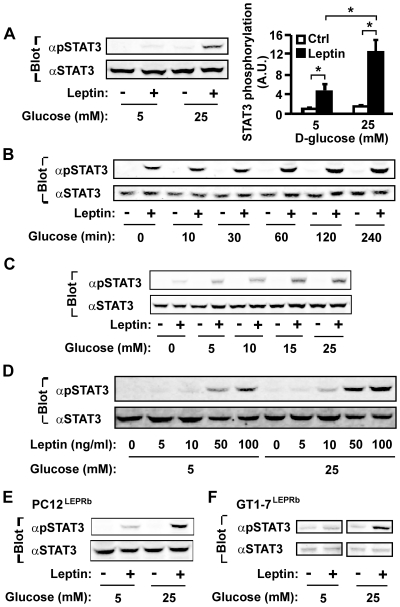
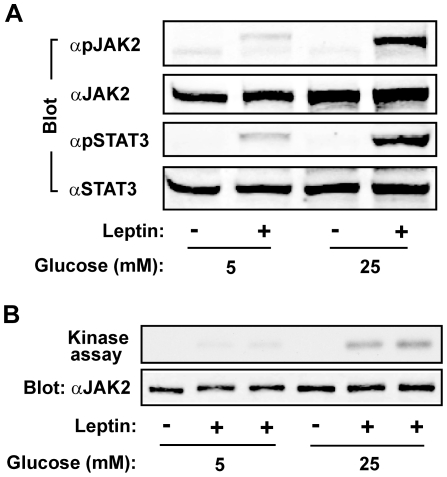
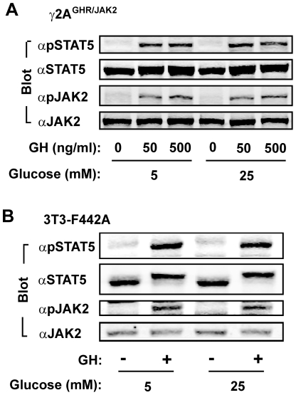
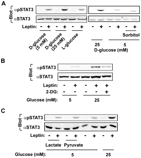
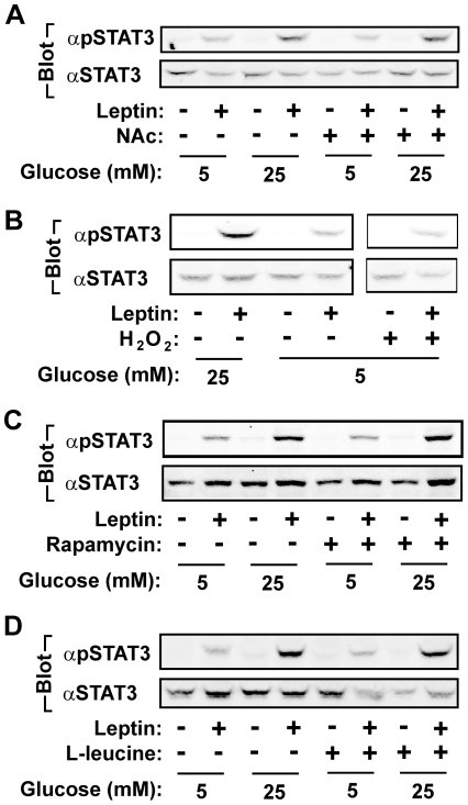
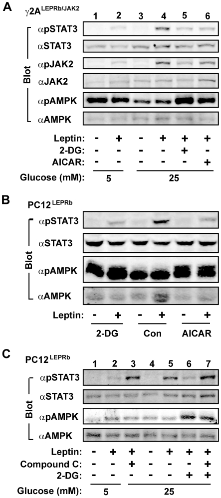
Leptin exerts its action by binding to and activating the long form of leptin receptors (LEPRb). LEPRb activates JAK2 that subsequently phosphorylates and activates STAT3. The JAK2/STAT3 pathway is required for leptin control of energy balance and body weight. Defects in leptin signaling lead to leptin resistance, a primary risk factor for obesity. Body weight is also regulated by nutrients, including glucose. Defects in glucose sensing also contribute to obesity. Here we report crosstalk between leptin and glucose. Glucose starvation blocked the ability of leptin to stimulate tyrosyl phosphorylation and activation of JAK2 and STAT3 in a variety of cell types. Glucose dose-dependently enhanced leptin signaling. In contrast, glucose did not enhance growth hormone-stimulated phosphorylation of JAK2 and STAT5. Glucose starvation or 2-deoxyglucose-induced inhibition of glycolysis activated AMPK and inhibited leptin signaling; pharmacological inhibition of AMPK restored the ability of leptin to stimulate STAT3 phosphorylation. Conversely, pharmacological activation of AMPK was sufficient to inhibit leptin signaling and to block the ability of glucose to enhance leptin signaling. These results suggest that glucose and/or its metabolites play a permissive role in leptin signaling, and that glucose enhances leptin sensitivity at least in part by attenuating the ability of AMPK to inhibit leptin signaling.
Conflict of interest statement
Figures
Similar articles
-
Leptin stimulates both JAK2-dependent and JAK2-independent signaling pathways.J Biol Chem. 2008 Oct 17;283(42):28066-73. doi: 10.1074/jbc.M805545200. Epub 2008 Aug 21. J Biol Chem. 2008. PMID: 18718905 Free PMC article.
-
Novel black soy peptides with antiobesity effects: activation of leptin-like signaling and AMP-activated protein kinase.Int J Obes (Lond). 2008 Jul;32(7):1161-70. doi: 10.1038/ijo.2008.60. Epub 2008 Apr 15. Int J Obes (Lond). 2008. PMID: 18414417
-
Leptin activates AMP-activated protein kinase in hepatic cells via a JAK2-dependent pathway.Biochem Biophys Res Commun. 2006 Dec 8;351(1):171-5. doi: 10.1016/j.bbrc.2006.10.015. Epub 2006 Oct 11. Biochem Biophys Res Commun. 2006. PMID: 17054914
-
Leptin receptor signaling and the regulation of mammalian physiology.Int J Obes (Lond). 2008 Dec;32 Suppl 7(Suppl 7):S8-12. doi: 10.1038/ijo.2008.232. Int J Obes (Lond). 2008. PMID: 19136996 Free PMC article. Review.
-
Leptin signaling in the hypothalamus: emphasis on energy homeostasis and leptin resistance.Front Neuroendocrinol. 2003 Dec;24(4):225-53. doi: 10.1016/j.yfrne.2003.10.001. Front Neuroendocrinol. 2003. PMID: 14726256 Review.
Cited by
-
Body Mass Index Influences the Salutary Effects of Metformin on Survival After Lobectomy for Stage I NSCLC.J Thorac Oncol. 2019 Dec;14(12):2181-2187. doi: 10.1016/j.jtho.2019.07.020. Epub 2019 Aug 6. J Thorac Oncol. 2019. PMID: 31398539 Free PMC article.
-
A twenty-first century cancer epidemic caused by obesity: the involvement of insulin, diabetes, and insulin-like growth factors.Int J Endocrinol. 2013;2013:632461. doi: 10.1155/2013/632461. Epub 2013 Jul 31. Int J Endocrinol. 2013. PMID: 23983688 Free PMC article.
-
Flurbiprofen Ameliorates Glucose Deprivation-Induced Leptin Resistance.Front Pharmacol. 2016 Sep 30;7:354. doi: 10.3389/fphar.2016.00354. eCollection 2016. Front Pharmacol. 2016. PMID: 27746736 Free PMC article.
-
Emerging role of leptin in rheumatoid arthritis.Clin Exp Immunol. 2014 Sep;177(3):557-70. doi: 10.1111/cei.12372. Clin Exp Immunol. 2014. PMID: 24802245 Free PMC article.
-
Lztfl1/BBS17 controls energy homeostasis by regulating the leptin signaling in the hypothalamic neurons.J Mol Cell Biol. 2018 Oct 1;10(5):402-410. doi: 10.1093/jmcb/mjy022. J Mol Cell Biol. 2018. PMID: 30423168 Free PMC article.
References
-
- Zhang Y, Proenca R, Maffei M, Barone M, Leopold L, et al. Positional cloning of the mouse obese gene and its human homologue. Nature. 1994;372:425–432. - PubMed
-
- Halaas JL, Gajiwala KS, Maffei M, Cohen SL, Chait BT, et al. Weight-reducing effects of the plasma protein encoded by the obese gene. Science. 1995;269:543–546. - PubMed
-
- Tartaglia LA, Dembski M, Weng X, Deng N, Culpepper J, et al. Identification and expression cloning of a leptin receptor, OB-R. Cell. 1995;83:1263–1271. - PubMed
-
- Chen H, Charlat O, Tartaglia LA, Woolf EA, Weng X, et al. Evidence that the diabetes gene encodes the leptin receptor: identification of a mutation in the leptin receptor gene in db/db mice. Cell. 1996;84:491–495. - PubMed
Publication types
MeSH terms
Substances
Grants and funding
LinkOut - more resources
Full Text Sources
Molecular Biology Databases
Miscellaneous
