Hemagglutinin from the H5N1 virus activates Janus kinase 3 to dysregulate innate immunity
- PMID: 22359619
- PMCID: PMC3280993
- DOI: 10.1371/journal.pone.0031721
Hemagglutinin from the H5N1 virus activates Janus kinase 3 to dysregulate innate immunity
Abstract
Highly pathogenic avian influenza viruses (HPAIVs) cause severe disease in humans. There are no effective vaccines or antiviral therapies currently available to control fatal outbreaks due in part to the lack of understanding of virus-mediated immunopathology. In our study, we used hemagglutinin (HA) of H5N1 virus to investigate the related signaling pathways and their relationship to dysregulated innate immune reaction. We found the HA of H5N1 avian influenza triggered an abnormal innate immune signalling in the pulmonary epithelial cells, through an unusual process involving activation of Janus kinase 3 (JAK3) that is exclusively associated with γc chain and is essential for signaling via all γc cytokine receptors. By using a selective JAK3 inhibitor and JAK3 knockout mice, we have, for the first time, demonstrated the ability to target active JAK3 to counteract injury to the lungs and protect immunocytes from acute hypercytokinemia -induced destruction following the challenge of H5N1 HA in vitro and in vivo. On the basis of the present data, it appears that the efficacy of selective JAK3 inhibition is likely based on its ability to block multiple cytokines and protect against a superinflammatory response to pathogen-associated molecular patterns (PAMPs) attack. Our findings highlight the potential value of selective JAK3 inhibitor in treating the fatal immunopathology caused by H5N1 challenge.
Conflict of interest statement
Figures


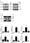
 , Jak3+/+ PBS,
  , Jak3−/− PBS,
  , Jak3+/+ HA,
  , Jak3−/− HA,
  , Jak3+/+ HA+JAK3Inh). *P<0.05 vs. Jak3+/+ PBS group; #
P<0.05 vs. Jak3+/+ HA group.


Similar articles
-
H5N1 Virus Hemagglutinin Inhibition of cAMP-Dependent CFTR via TLR4-Mediated Janus Tyrosine Kinase 3 Activation Exacerbates Lung Inflammation.Mol Med. 2015 Jan 12;21(1):134-42. doi: 10.2119/molmed.2014.00189. Mol Med. 2015. PMID: 25587856 Free PMC article.
-
A novel hemagglutinin protein produced in bacteria protects chickens against H5N1 highly pathogenic avian influenza viruses by inducing H5 subtype-specific neutralizing antibodies.PLoS One. 2017 Feb 17;12(2):e0172008. doi: 10.1371/journal.pone.0172008. eCollection 2017. PLoS One. 2017. PMID: 28212428 Free PMC article.
-
Recombinant modified vaccinia virus Ankara expressing the hemagglutinin gene confers protection against homologous and heterologous H5N1 influenza virus infections in macaques.J Infect Dis. 2009 Feb 1;199(3):405-13. doi: 10.1086/595984. J Infect Dis. 2009. PMID: 19061423
-
[Cytokine storm in avian influenza].Mikrobiyol Bul. 2008 Apr;42(2):365-80. Mikrobiyol Bul. 2008. PMID: 18697437 Review. Turkish.
-
[Tetravaccine--new fundamental approach to prevention of influenza pandemic].Zh Mikrobiol Epidemiol Immunobiol. 2007 Jul-Aug;(4):15-9. Zh Mikrobiol Epidemiol Immunobiol. 2007. PMID: 17882832 Review. Russian.
Cited by
-
H5N1 Virus Hemagglutinin Inhibition of cAMP-Dependent CFTR via TLR4-Mediated Janus Tyrosine Kinase 3 Activation Exacerbates Lung Inflammation.Mol Med. 2015 Jan 12;21(1):134-42. doi: 10.2119/molmed.2014.00189. Mol Med. 2015. PMID: 25587856 Free PMC article.
-
P2Y6 receptors are involved in mediating the effect of inactivated avian influenza virus H5N1 on IL-6 & CXCL8 mRNA expression in respiratory epithelium.PLoS One. 2017 May 11;12(5):e0176974. doi: 10.1371/journal.pone.0176974. eCollection 2017. PLoS One. 2017. PMID: 28494003 Free PMC article.
-
Network-Based Integrated Analysis of Transcriptomic Studies in Dissecting Gene Signatures for LPS-Induced Acute Lung Injury.Inflammation. 2021 Dec;44(6):2486-2498. doi: 10.1007/s10753-021-01518-8. Epub 2021 Aug 30. Inflammation. 2021. PMID: 34462829 Free PMC article.
-
Role of miRNA in Highly Pathogenic H5 Avian Influenza Virus Infection: An Emphasis on Cellular and Chicken Models.Viruses. 2024 Jul 9;16(7):1102. doi: 10.3390/v16071102. Viruses. 2024. PMID: 39066264 Free PMC article. Review.
-
Host transcriptome-guided drug repurposing for COVID-19 treatment: a meta-analysis based approach.PeerJ. 2020 Jun 10;8:e9357. doi: 10.7717/peerj.9357. eCollection 2020. PeerJ. 2020. PMID: 32566414 Free PMC article.
References
-
- Swayne DE, Suarez DL. Highly pathogenic avian influenza. Rev Sci Tech. 2000;19:463–482. - PubMed
-
- WHO. 2010. Cumulative Number of Confirmed Human Cases of Avian Influenza A/(H5N1) Reported to WHO.
Publication types
MeSH terms
Substances
Grants and funding
LinkOut - more resources
Full Text Sources
Other Literature Sources
Medical
Molecular Biology Databases

