. 2012;7(2):e31807.
doi: 10.1371/journal.pone.0031807. Epub 2012 Feb 16.

L-cysteine administration attenuates pancreatic fibrosis induced by TNBS in rats by inhibiting the activation of pancreatic stellate cell

Affiliations

L-cysteine administration attenuates pancreatic fibrosis induced by TNBS in rats by inhibiting the activation of pancreatic stellate cell

LiJuan Yang et al. PLoS One. 2012.

Abstract

Background and aims: Recent studies have shown that activated pancreatic stellate cells (PSCs) play a major role in pancreatic fibrogenesis. We aimed to study the effect of L-cysteine administration on fibrosis in chronic pancreatitis (CP) induced by trinitrobenzene sulfonic acid (TNBS) in rats and on the function of cultured PSCs.

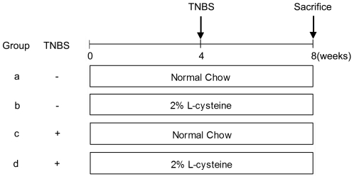
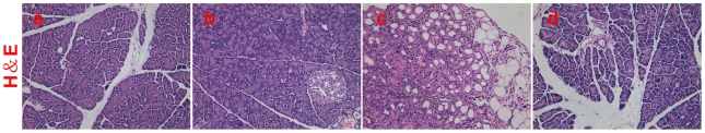
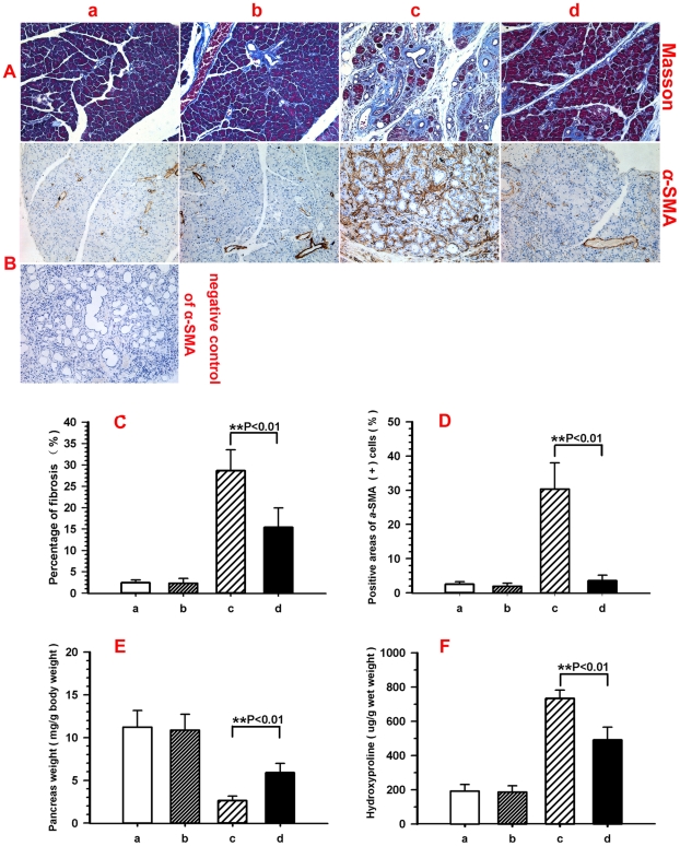
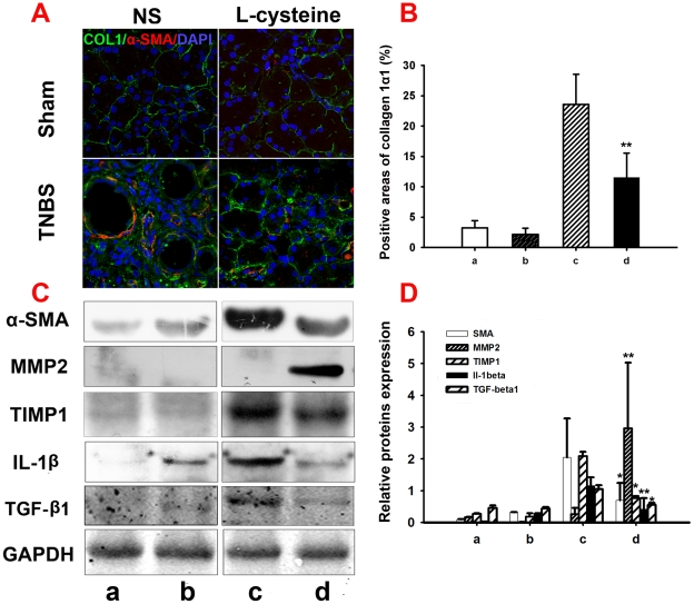
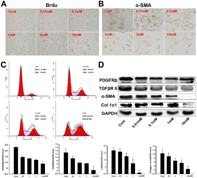
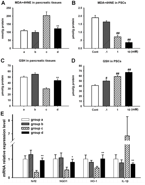
Methods: CP was induced by TNBS infusion into rat pancreatic ducts. L-cysteine was administrated for the duration of the experiment. Histological analysis and the contents of hydroxyproline were used to evaluate pancreatic damage and fibrosis. Immunohistochemical analysis of α-SMA in the pancreas was performed to detect the activation of PSCs in vivo. The collagen deposition related proteins and cytokines were determined by western blot analysis. DNA synthesis of cultured PSCs was evaluated by BrdU incorporation. We also evaluated the effect of L-cysteine on the cell cycle and cell activation by flow cytometry and immunocytochemistry. The expression of PDGFRβ, TGFβRII, collagen 1α1 and α-SMA of PSCs treated with different concentrations of L-cysteine was determined by western blot. Parameters of oxidant stress were evaluated in vitro and in vivo. Nrf2, NQO1, HO-1, IL-1β expression were evaluated in pancreas tissues by qRT-PCR.

Results: The inhibition of pancreatic fibrosis by L-cysteine was confirmed by histological observation and hydroxyproline assay. α-SMA, TIMP1, IL-1β and TGF-β1 production decreased compared with the untreated group along with an increase in MMP2 production. L-cysteine suppressed the proliferation and extracellular matrix production of PSCs through down-regulating of PDGFRβ and TGFβRII. Concentrations of MDA+4-HNE were decreased by L-cysteine administration along with an increase in GSH levels both in tissues and cells. In addition, L-cysteine increased the mRNA expression of Nrf2, NQO1 and HO-1 and reduced the expression of IL-1β in L-cysteine treated group when compared with control group.

Conclusion: L-cysteine treatment attenuated pancreatic fibrosis in chronic pancreatitis in rats.


Conflict of interest statement

Competing Interests: The authors have declared that no competing interests exist.

Figures

Figure 1
Figure 1. Schematic map of the experimental design.
Four groups of rats (n = 10) were studied. Groups a and b received saline injections (no induction of chronic pancreatitis), groups c and d received TNBS injections (0.4 ml of 2% TNBS for the induction of chronic pancreatitis). Groups a and c received normal chow throughout the entire 8-week study period. Groups b and d received chow mixed with 2% L-cysteine during the 8-week study, after which the rats were sacrificed. Arrows indicate injections with TNBS.
Figure 2
Figure 2. Histological observations in H&E stained sections.
For the description of groups a to d, see Figure 1. Abnormal architecture, glandular atrophy, pseudotubular complexes, fibrosis and inflammatory cell infiltration can be seen in group c, while appearance is fairly normal in groups a and b. Representative H&E sections of rats revealed that 2% L-cysteine attenuated the development of pancreatic fibrosis induced by TNBS (group d). Representative original magnification ×200.
Figure 3
Figure 3. L-cysteine attenuates pancreatic fibrosis induced by TNBS.
For the description of groups a to d, see Figure 1. (A, B) Pancreatic tissue sections stained with Masson and stained immunohistochemically for α-SMA. (C, D) Quantification of the positive areas of Masson and α-SMA by Image Pro Plus. (E) Effect of L-cysteine treatment on pancreas wet weight. Pancreas wet weight was significantly increased in L-cysteine treated rats. (F) Effect of L-cysteine treatment on pancreatic content of hydroxyproline after TNBS injury. Values are mean±SD (n = 10). Significant differences: **p<0.01 group c vs. group d. Representative original magnification ×200.
Figure 4
Figure 4. L-cysteine modulates extracellular matrix secretion in vitro.
For the description of groups a to d, see Figure 1. (A, B) Double immunofluorescence of collagen 1α1 (green) and α-SMA (red) in the pancreas, 4′, 6-Diamidino-2-phenylindole (DAPI; blue) was used to counterstain nuclei. The co-localization of collagen 1α1 and α-SMA is highlighted by the yellow color. Immunostainning showed a low expression of collagen 1α1 in the sham pancreas, but its expression increased obviously after 4 weeks TNBS treatment, while L-cysteine administration attenuated collagen 1α1 expression in CP rats. (C, D) Expression of α-SMA, MMP2, TIMP1, TGF-β1 and IL-1β proteins in 4 groups of pancreatic tissues were detected by western blot analysis. GAPDH was used as the loading control in all experiments. The results were quantified by determining the intensities of the bands compared with those of GAPDH. All data are presented as the mean±SD of three independent experiments. Significant differences: *p<0.05 vs. group c, **p<0.01 vs. group c. Representative original magnification ×400.
Figure 5
Figure 5. L-cysteine inhibits the proliferation and activation of PSCs.
(A) Brdu staining of PSCs three days after L-cysteine treatment, DNA synthesis was detected by BrdU incorporation during the final 2 hours (dividing cells are stained dark brown). (B) Effect of L-cysteine on the activation of freshly isolated PSCs. After culturing PSCs isolated from a rat for 24 hour, the medium was changed to MEM+0–10 mM L-cysteine. PSCs were cultured for five days under the above conditions and α-SMA expression by PSCs was measured with immunocytochemistry. (C) The cell cycle distribution of PSCs at different concentrations of L-cysteine was analyzed by flow cytometry. (D) Expression of PDGFRβ, TGFβRII, α-SMA and collagen 1α1 protein in the PSCs treated with different concentrations of L-cysteine was analyzed by western blot. GAPDH was used as a loading control. The intensities of bands were measured, and the represented values correspond to the PDGFRβ, TGFβRII, α-SMA and collagen 1α1/GAPDH ratio. All data are presented as the mean±SD of three independent experiments. Significant differences: *p<0.05 vs. control group, **p<0.01 vs. control group. Representative original magnification ×200.
Figure 6
Figure 6. L-cysteine modulates oxidative stress in vitro and in vivo and regulates Nrf2 associated pathway.
For the description of groups a to d, see Figure 1. (A) MDA+4HNE concentration of pancreatic tissues. MDA+4HNE concentration of pancreatic tissues increased significantly after induction of CP compared with sham groups and decreased obviously after L-cysteine administration. (C) GSH levels of pancreatic tissues. GSH levels of pancreatic tissues decrease significantly after TNBS administration compared with sham groups and increased obviously after L-cysteine administration. (B, D) In vitro study, L-cysteine treatment affected MDA+4-HNE and GSH levels in a dose-dependent manner in PSCs as that in in vivo study. (E) Relative mRNA levels of Nrf2, NQO1, HO-1 and IL-1β after L-cysteine treatment in pancreas of four groups. All data are presented as the mean±SD of three independent experiments. Significant differences: *p<0.05 vs. group c, **p<0.01 vs. group c, # p<0.05 vs. control group, ## p<0.01 vs. control group.

References

    1. Apte MV, Wilson JS. Mechanisms of pancreatic fibrosis. Dig Dis. 2004;22:272–279. - PubMed
    1. Clain JE, Pearson RK. Diagnosis of chronic pancreatitis: is a gold standard necessary? Surg Clin North Am. 1999;79:829–845. - PubMed
    1. Khalid A, Whitcomb DC. Conservative management of chronic pancreatitis. Eur J Gastroenterol Hepatol. 2002;14:943–949. - PubMed
    1. Laugier R, Grandval P. Interventional treatment of chronic pancreatitis. Eur J Gastroenterol Hepatol. 2002;14:951–955. - PubMed
    1. Levy MJ, Wiersema MJ. EUS-guided celiac plexus neurolysis and celiac plexus block. Gastrointest Endosc. 2003;57:923–930. - PubMed

Publication types