

Nonadhesive culture system as a model of rapid sphere formation with cancer stem cell properties
- PMID: 22359637
- PMCID: PMC3281010
- DOI: 10.1371/journal.pone.0031864
Nonadhesive culture system as a model of rapid sphere formation with cancer stem cell properties
Abstract
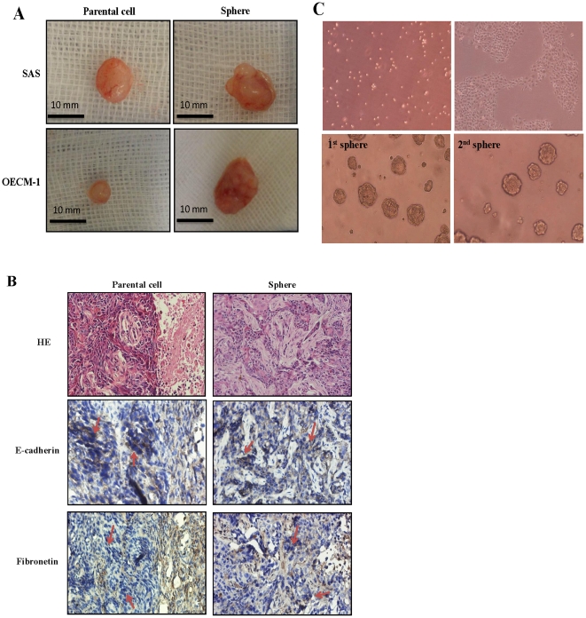
Background: Cancer stem cells (CSCs) play an important role in tumor initiation, progression, and metastasis and are responsible for high therapeutic failure rates. Identification and characterization of CSC are crucial for facilitating the monitoring, therapy, or prevention of cancer. Great efforts have been paid to develop a more effective methodology. Nevertheless, the ideal model for CSC research is still evolving. In this study, we created a nonadhesive culture system to enrich CSCs from human oral squamous cell carcinoma cell lines with sphere formation and to characterize their CSC properties further.
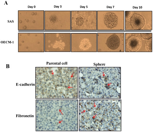
Methods: A nonadhesive culture system was designed to generate spheres from the SAS and OECM-1 cell lines. A subsequent investigation of their CSC properties, including stemness, self-renewal, and chemo- and radioresistance in vitro, as well as tumor initiation capacity in vivo, was also performed.
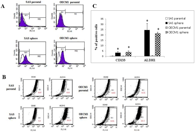
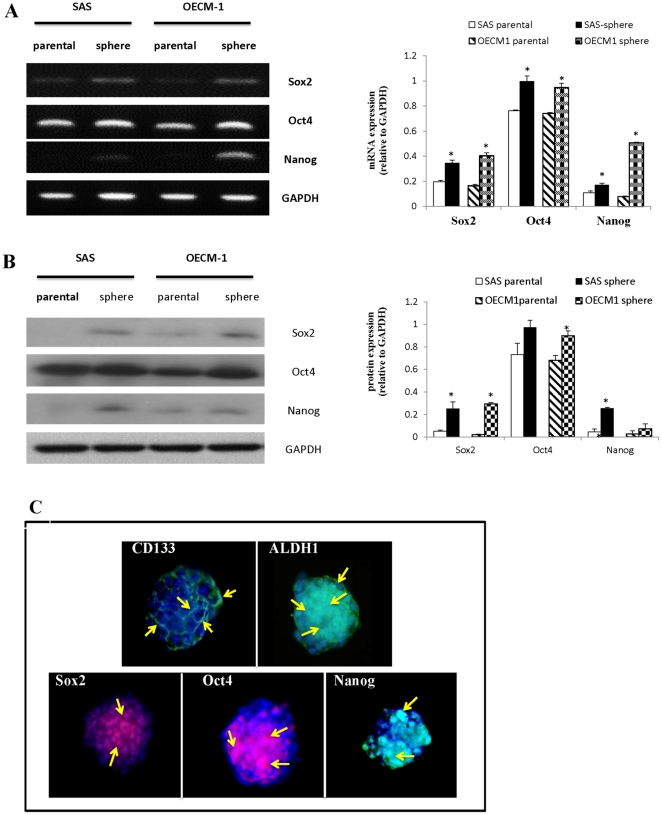
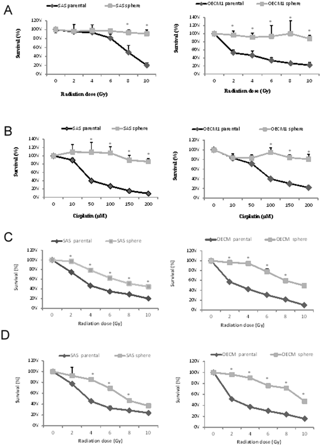
Results: Spheres were formed cost-effectively and time-efficiently within 5 to 7 days. Moreover, we proved that these spheres expressed putative stem cell markers and exhibited chemoradiotherapeutic resistance, in addition to tumor-initiating and self-renewal capabilities.
Conclusions: Using this nonadhesive culture system, we successfully established a rapid and cost-effective model that exhibits the characteristics of CSCs and can be used in cancer research.
Conflict of interest statement
Figures
Similar articles
-
Enrichment and characterization of cancer stem‑like cells from a cervical cancer cell line.Mol Med Rep. 2014 Jun;9(6):2117-23. doi: 10.3892/mmr.2014.2063. Epub 2014 Mar 20. Mol Med Rep. 2014. PMID: 24676900 Free PMC article.
-
Sphere-forming cell subpopulations with cancer stem cell properties in human hepatoma cell lines.BMC Gastroenterol. 2011 Jun 14;11:71. doi: 10.1186/1471-230X-11-71. BMC Gastroenterol. 2011. PMID: 21669008 Free PMC article.
-
Enrichment and characterization of cancer stem-like cells in ultra-low concentration of serum and non-adhesive culture system.Am J Transl Res. 2018 May 15;10(5):1552-1561. eCollection 2018. Am J Transl Res. 2018. PMID: 29887968 Free PMC article.
-
Tumor-derived spheroids: Relevance to cancer stem cells and clinical applications.Cancer Sci. 2017 Mar;108(3):283-289. doi: 10.1111/cas.13155. Cancer Sci. 2017. PMID: 28064442 Free PMC article. Review.
-
Cancer stem cells - new and potentially important targets for the therapy of oral squamous cell carcinoma.Oral Dis. 2006 Sep;12(5):443-54. doi: 10.1111/j.1601-0825.2006.01264.x. Oral Dis. 2006. PMID: 16910914 Review.
Cited by
-
Quercetin suppresses drug-resistant spheres via the p38 MAPK-Hsp27 apoptotic pathway in oral cancer cells.PLoS One. 2012;7(11):e49275. doi: 10.1371/journal.pone.0049275. Epub 2012 Nov 12. PLoS One. 2012. PMID: 23152886 Free PMC article.
-
The ULK3 kinase is a determinant of keratinocyte self-renewal and tumorigenesis targeting the arginine methylome.Nat Commun. 2023 Feb 16;14(1):887. doi: 10.1038/s41467-023-36410-6. Nat Commun. 2023. PMID: 36797248 Free PMC article.
-
Cancer-Associated Fibroblasts Promote Tumor Aggressiveness in Head and Neck Cancer through Chemokine Ligand 11 and C-C Motif Chemokine Receptor 3 Signaling Circuit.Cancers (Basel). 2022 Jun 27;14(13):3141. doi: 10.3390/cancers14133141. Cancers (Basel). 2022. PMID: 35804913 Free PMC article.
-
Transcriptomic study of the mechanism of anoikis resistance in head and neck squamous carcinoma.PeerJ. 2019 May 23;7:e6978. doi: 10.7717/peerj.6978. eCollection 2019. PeerJ. 2019. PMID: 31198634 Free PMC article.
-
A site-specific phosphorylation of the focal adhesion kinase controls the formation of spheroid cell clusters.Neurochem Res. 2014 Jul;39(7):1199-205. doi: 10.1007/s11064-014-1298-y. Epub 2014 Apr 5. Neurochem Res. 2014. PMID: 24706070
References
-
- Jemal A, Bray F, Center MM, Ferlay J, Ward E, et al. Global cancer statistics. CA Cancer J Clin. 2011;61:69–90. - PubMed
-
- Al-Swiahb JN, Chen CH, Chuang HC, Fang FM, Tasi HT, et al. Cinical, pathological and molecular determinants in squamous cell carcinoma of the oral cavity. Future Oncol. 2010;6:837–850. - PubMed
-
- Olasz L, Szabo I, Horvath A. A combined treatment for advanced oral cavity cancers. Cancer. 1988;62:1267–1274. - PubMed
-
- Lippman SM, Sudbo J, Hong WK. Oral cancer prevention and the evolution of molecular-targeted drug development. J Clin Oncol. 2005;23:346–356. - PubMed
-
- Le Tourneau C. Molecularly targeted therapy in head and neck cancer. Bull Cancer. 2010;97:1453–1466. - PubMed
Publication types
MeSH terms
LinkOut - more resources
Full Text Sources
Other Literature Sources
