Review
. 2012 May;53(5):779-89.
doi: 10.1111/j.1528-1167.2012.03421.x. Epub 2012 Feb 23.

A brief history on the oscillating roles of thalamus and cortex in absence seizures

Affiliations
Review

A brief history on the oscillating roles of thalamus and cortex in absence seizures

Massimo Avoli. Epilepsia. 2012 May.

Abstract

This review summarizes the findings obtained over the past 70 years on the fundamental mechanisms underlying generalized spike-wave (SW) discharges associated with absence seizures. Thalamus and cerebral cortex are the brain areas that have attracted most of the attention from both clinical and experimental researchers. However, these studies have often favored either one or the other structure in playing a major role, thus leading to conflicting interpretations. Beginning with Jasper and Penfield's topistic view of absence seizures as the result of abnormal functions in the so-called centrencephalon, we witness the naissance of a broader concept that considered both thalamus and cortex as equal players in the process of SW discharge generation. Furthermore, we discuss how recent studies have identified fine changes in cortical and thalamic excitability that may account for the expression of absence seizures in naturally occurring genetic rodent models and knockout mice. The end of this fascinating tale is presumably far from being written. However, I can confidently conclude that in the unfolding of this "novel," we have discovered several molecular, cellular, and pharmacologic mechanisms that govern forebrain excitability, and thus consciousness, during the awake state and sleep.


Conflict of interest statement

Disclosure

I have no conflict of interest to disclose. I also confirm that I have read the Journal’s position on issues involved in ethical publication and I affirm that this report is consistent with those guidelines.

Figures
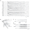
Figure 1
Figure 1
(A) Generalized 3-Hz SW discharges recorded from a 10-year-old boy with childhood absence epilepsy. Note the generalized onset of the discharge, its synchronization between the two hemispheres, and its sudden termination that occurs simultaneously in all brain regions. EEG recording kindly provided by Drs. Francesca Pittau, Jean Gotman, and Francois Dubeau at the Montreal Neurological Institute & Hospital. (B) Drawing of the hypothetical organization of the thalamocortical systems showing the direct specific relay system (R), the specific association system (A), as well as the superimposed polineuronal thalamic reticular system. Modified from Jasper (1949). (C) Bilaterally synchronous SW pattern produced in the cat cortex by 3-Hz stimulation of the thalamic massa intermedia. Modified from EEG recordings as in Jasper and Droogleever-Fortuyn (1946). (D) Generalized SW discharges induced by bilateral application of cobalt to the frontal areas of a rhesus monkey; these seizures were associated with absences and, at times, with single myoclonic jerks. Bipolar EEG recordings were obtained from the prefrontal (PF), premotor (PM), and precentral regions. Modified from EEG recordings as in Marcus and Watson (1968). Epilepsia © ILAE
Figure 2
Figure 2
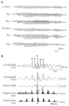
(A) Generalized SW discharges recorded in a cat following intramuscular injection of a large dose of penicillin. Note the EEG similarities (but for the higher frequency) between cat and human recordings as shown in Fig. 1A. (B) EEG averages and single-unit perievent histograms triggered by the negative peaks of the spikes of SW discharges induced by penicillin injection and recorded intracortically (dots in the upper trace). The cortical unit was recorded in the middle suprasylvian gyrus, whereas the thalamic unit was recorded simultaneously from the nucleus lateralis posterior/pulvinar complex. Note the late involvement of the thalamic unit in SW firing as well as the two peaks of firing probability, one (straight arrow) preceding, and the other (curved arrow) coinciding, with the cortical peak of firing probability. The thalamic unit recorded in this experiment was orthodromically activated by electrical stimuli delivered in the cortex middle suprasylvian gyrus (not illustrated). Epilepsia © ILAE
Figure 3
Figure 3
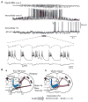
(A) Drawing of an intracellular signal recorded from a thalamocortical relay neuron that was hyperpolarized with injection of a square pulse of negative current (arrows point at the onset and termination of the hyperpolarizing command). Note the “sag” of the membrane that leads over time to less polarized values as well as the burst of action potentials generated upon termination of the pulse. These two phenomena are known to be caused by Ih and by T-type Ca2+ current, respectively. (B) Schematic diagram of the thalamocortical loop. Glutamatergic cortical and thalamocortical relay cells are shown in red, whereas reticular nucleus GABAergic interneurons are colored in blue. (C) Drawing of the intracellular activity generated by a thalamocortical relay neuron during a “sleep” spindle. Each cycle of the spindle begins with a hyperpolarizing IPSP that deinactivates the T-type Ca2+ current and causes a progressive activation of Ih. As the membrane becomes less polarized a low threshold Ca2+ spike, which can cause action potential bursting, is generated; these action potentials will excite reticular nucleus GABAergic cells which, in turn, will cause a hyperpolarizing IPSP in thalamocortical relay cells thus starting another spindle cycle. (D) Intracellular, spindle oscillations recorded in vitro from a perigeniculate (GABAergic) and geniculate (relay) neuron under control conditions and during blockade of GABAA receptors (bicuculline) (Modified from Bal et al., 1995a). Epilepsia © ILAE
Figure 4
Figure 4
(A) SW discharge recorded intracortically (depth-EEG area 4) and simultaneously with dual intracellular recordings from a cortical (Intracellular area 4) and a thalamocortical (intracellular VL) neuron in a cat under ketamine–xylazine anesthesia. The portion indicated by the thick line is expanded below. Note the progressive steady depolarization and concomitant action potential bursting in the cortical neuron while phasic IPSPs (arrows) occur in the thalamocortical cell in phase with the cortical excitatory events. Note, also, that the brief period of quiescence in cortical discharge coincides with action potential firing in the thalamocortical cell (arrow) (modified from unpublished data by M. Steriade and D. Contreras). (B) Simplified diagrammatic summary of the results obtained by Meeren et al. (2002) by employing nonlinear association analysis of the EEG signals recorded simultaneously from multiple cortical and thalamic structures during spontaneous SW discharges in WAG/Rij rats. Corticocortical (black arrows), intrathalamic (blue arrows), and thalamocorticothalamic (red arrows) interdependencies are shown during the first 500 msec of the SW discharge (a) and for the entire duration of the SW discharge (b). The thickness of the arrow represents the average strength of the association, and the direction of the arrowhead points to the direction of the lagging site. Note that SW discharge initiates in the upper lip/nose area of the somatosensory cortex and then propagates to other cortical regions and to thalamus, mainly to its laterodorsal nucleus. When the entire seizure is analyzed as one epoch, the same cortical initiation site (“focus”) as during the first 500 msec is found consistently but, compared with the first 500 msec, the strength of association between ventroposterior lateral (VPL) and ventroposterior medial (VPM) nuclei has increased, and the direction of the thalamocorticothalamic couplings has changed. Epilepsia © ILAE

References

    1. Avanzini G, Vergnes M, Spreafico R, Marescaux C. Calcium-dependent regulation of genetically determined spike and waves by the reticular thalamic nucleus of rats. Epilepsia. 1993;34:1–7. - PubMed
    1. Bal T, von Krosigk M, McCormick DA. Synaptic and membrane mechanisms underlying synchronized oscillations in the ferret lateral geniculate nucleus in vitro. J Physiol. 1995a;483:641–663. - PMC - PubMed
    1. Bal T, von Krosigk M, McCormick DA. Role of the ferret perigeniculate nucleus in the generation of synchronized oscillations in vitro. J Physiol. 1995b;483:665–685. - PMC - PubMed
    1. Bal T, Debay D, Destexhe A. Cortical feedback controls the frequency and synchrony of oscillations in the visual thalamus. J Neurosci. 2000;20:7478–7488. - PMC - PubMed
    1. Bancaud J, Talairach J, Morel P, Bresson M, Bonis A, Geier S, Hemon E, Buser P. “Generalized” epileptic seizures elicited by electrical stimulation of the frontal lobe in man. Electroencephalogr Clin Neurophysiol. 1974;37:275–282. - PubMed

Grants and funding