. 2012 May 18;7(5):863-70.
doi: 10.1021/cb200311s. Epub 2012 Feb 24.

Sulfolipid-1 biosynthesis restricts Mycobacterium tuberculosis growth in human macrophages

Affiliations
Free PMC article

Sulfolipid-1 biosynthesis restricts Mycobacterium tuberculosis growth in human macrophages

Sarah A Gilmore et al. ACS Chem Biol. .
Free PMC article

Abstract

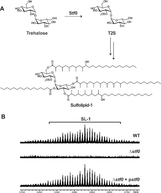
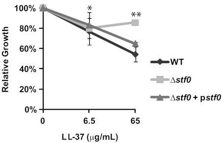
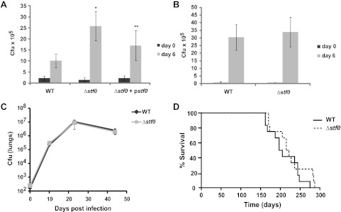
Mycobacterium tuberculosis (Mtb), the causative agent of tuberculosis, is a highly evolved human pathogen characterized by its formidable cell wall. Many unique lipids and glycolipids from the Mtb cell wall are thought to be virulence factors that mediate host-pathogen interactions. An intriguing example is Sulfolipid-1 (SL-1), a sulfated glycolipid that has been implicated in Mtb pathogenesis, although no direct role for SL-1 in virulence has been established. Previously, we described the biochemical activity of the sulfotransferase Stf0 that initiates SL-1 biosynthesis. Here we show that a stf0-deletion mutant exhibits augmented survival in human but not murine macrophages, suggesting that SL-1 negatively regulates the intracellular growth of Mtb in a species-specific manner. Furthermore, we demonstrate that SL-1 plays a role in mediating the susceptibility of Mtb to a human cationic antimicrobial peptide in vitro, despite being dispensable for maintaining overall cell envelope integrity. Thus, we hypothesize that the species-specific phenotype of the stf0 mutant is reflective of differences in antimycobacterial effector mechanisms of macrophages.


Figures

Figure 1
Figure 1
Structure of SL-1 and a synthetic analogue. The synthetic analogue of SL-1 is composed of a T2S core esterified with fatty acyl groups at the 2′, 3′, 6′, and 6 positions, similar to SL-1. However, due to the complex synthesis of the fatty acyl substituents found on the natural product, SL-A lacks the hydroxyl groups and multimethyl branched units found on SL-1.
Figure 2
Figure 2
SL-1 induces a unique transcriptional signature in human leukocytes. The transcript profiles of hiDCs stimulated with P3K, SL-1, SL-A, or vehicle only were assessed by microarray. The 50 most highly upregulated and statistically significant transcripts (p < 0.02) induced by SL-1, SL-A, or P3K as compared to vehicle only treated cells were organized according to function. Due to limited overlap in genes upregulated by each stimulus, different functional categories were chosen. The percent overlap of gene identities between P3K vs SL-1, P3K vs SL-A, and SL-1 vs SL-A is highlighted. The identities of individual genes are listed in Supplementary Table 1.
Figure 3
Figure 3
Sulfation of trehalose by Stf0 is the first committed step in SL-1 biosynthesis. (A) The sulfotransferase Stf0 sulfates free trehalose to initiate SL-1 biosynthesis. (B) SL-1 is absent in the Δstf0 strain. Surface exposed lipids were removed by hexanes extraction of WT, Δstf0, and complemented (Δstf0 + pstf0) Mtb strains and analyzed by FT-ICR MS. SL-1 is observed as a collection of lipoforms that vary in the lengths of their acyl groups (±14 mass units).
Figure 4
Figure 4
Δstf0 is more resistant to LL-37 compared to WT Mtb. The indicated strains of Mtb were exposed to increasing concentrations (0, 6.5, and 65 μg/mL) of LL-37. After 3 days, Mtb viability was measured by plating bacteria on solid agar to enumerate cfu counts. Data are representative of one of three independent experiments each performed in triplicate. * P = 0.74 (not significant) for the comparison of Δstf0 versus WT; ** P = 0.012 for the comparison of Δstf0 versus WT. Error bars correspond to the means (±SD); an error of less than ∼2% is not visible on this graph (SD of Δstf0 at 65 μg/mL = 1.0%; SD of Δstf0 + pstf0 at 65 μg/mL = 1.27%).
Figure 5
Figure 5
Stf0 restricts the intracellular growth of Mtb in human macrophages. (A) Human THP-1 macrophages or (B) murine RAW macrophages were infected with the indicated strain of Mtb. After 4 h (day 0) and 6 days post infection, adherent macrophages were lysed and plated on solid agar to determine the number of viable intracellular bacteria by cfu counts. Data represent the average cfu count obtained from three independent experiments performed in triplicate (±SD). * P < 4 × 10⁻⁵ for the comparison of Δstf0 versus WT; ** P < 0.03 for the comparison of Δstf0 versus Δstf0 + pstf0 (A) or one of three independent experiments performed in triplicate (B) (±SD). Δstf0 behaves similarly to WT Mtb in a murine model of infection. (C) Lung cfu counts for BALB/c mice infected via aerosol with WT or Δstf0 Mtb. Each data point represents the average cfu count from 4–5 mice, and error bars indicate the standard deviation from the mean. (D) Time-to-death for mice infected with WT or Δstf0 Mtb.

Similar articles

Cited by

References

    1. Kaur D.; Guerin M. E.; Skovierova H.; Brennan P. J.; Jackson M. (2009) Chapter 2: Biogenesis of the cell wall and other glycoconjugates of Mycobacterium tuberculosis. Adv. Appl. Microbiol. 69, 23–78. - PMC - PubMed
    1. Reed M. B.; Domenech P.; Manca C.; Su H.; Barczak A. K.; Kreiswirth B. N.; Kaplan G.; Barry C. E. 3rd. (2004) A glycolipid of hypervirulent tuberculosis strains that inhibits the innate immune response. Nature 431, 84–87. - PubMed
    1. Geijtenbeek T. B.; Van Vliet S. J.; Koppel E. A.; Sanchez-Hernandez M.; Vandenbroucke-Grauls C. M.; Appelmelk B.; Van Kooyk Y. (2003) Mycobacteria target DC-SIGN to suppress dendritic cell function. J. Exp. Med. 197, 7–17. - PMC - PubMed
    1. Bowdish D. M.; Sakamoto K.; Kim M. J.; Kroos M.; Mukhopadhyay S.; Leifer C. A.; Tryggvason K.; Gordon S.; Russell D. G. (2009) MARCO, TLR2, and CD14 are required for macrophage cytokine responses to mycobacterial trehalose dimycolate and Mycobacterium tuberculosis. PLoS Pathog. 5, e1000474. - PMC - PubMed
    1. Cole S. T.; Brosch R.; Parkhill J.; Garnier T.; Churcher C.; Harris D.; Gordon S. V.; Eiglmeier K.; Gas S.; Barry C. E. 3rd; Tekaia F.; Badcock K.; Basham D.; Brown D.; Chillingworth T.; Connor R.; Davies R.; Devlin K.; Feltwell T.; Gentles S.; Hamlin N.; Holroyd S.; Hornsby T.; Jagels K.; Barrell B. G.; Krogh A; Mclean J; Moule S; Murphy L; Oliver K; Osborne J; Quail M. A.; Rajandream M. A.; Rogers J; Rutter S; Seeger K; Skelton J; Squares R; Squares S; Sulston J. E.; Taylor K; Whitehead S; Barrell B. G. (1998) Deciphering the biology of Mycobacterium tuberculosis from the complete genome sequence. Nature 393, 537–544. - PubMed

Publication types