. 2012 Sep 15;17(6):849-59.
doi: 10.1089/ars.2011.4507. Epub 2012 Apr 20.

The neuroprotection of hydrogen sulfide against MPTP-induced dopaminergic neuron degeneration involves uncoupling protein 2 rather than ATP-sensitive potassium channels

Affiliations

The neuroprotection of hydrogen sulfide against MPTP-induced dopaminergic neuron degeneration involves uncoupling protein 2 rather than ATP-sensitive potassium channels

Ming Lu et al. Antioxid Redox Signal. .

Abstract

Aims: Hydrogen sulfide (H(2)S), a novel gaseous mediator, has been recognized to protect neurons from overexcitation by enhancing the activity of the adenosine triphosphate-sensitive potassium (K-ATP) channel. However, no direct evidence supports that the K-ATP channel contributes to the neuroprotective effect of H(2)S in neurodegeneration. Herein, wild-type and Kir6.2 knockout (Kir6.2(-/-)) mice were used to establish the 1-methyl-4-phenyl-1,2,3,6-tetrahydropyridine (MPTP) mouse model of Parkinson's disease (PD) so as to investigate the involvement of K-ATP channels in the neuroprotection of H(2)S.

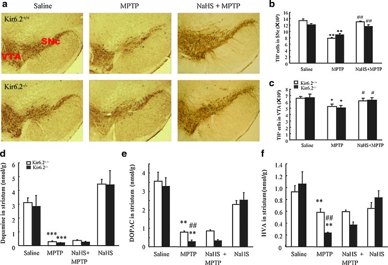
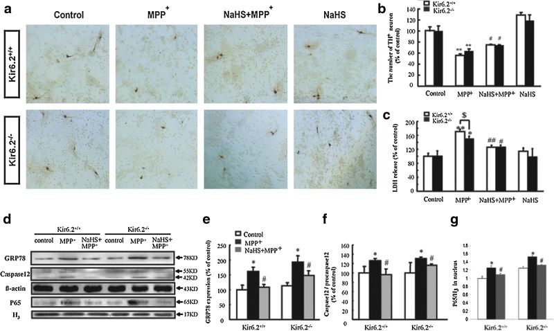
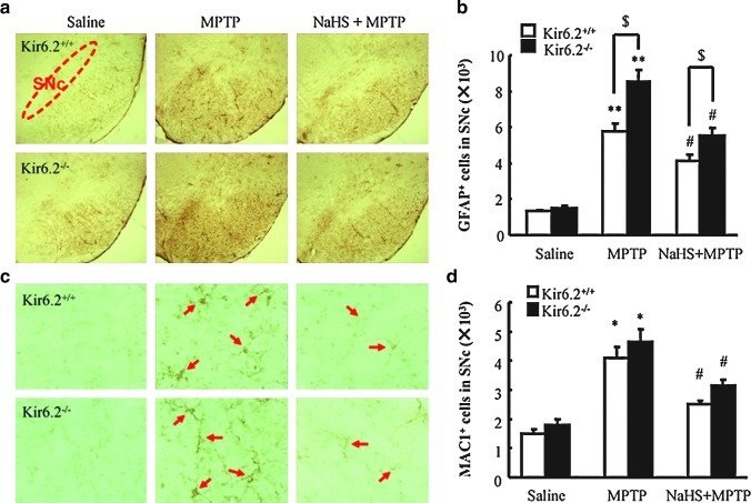
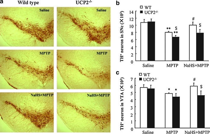
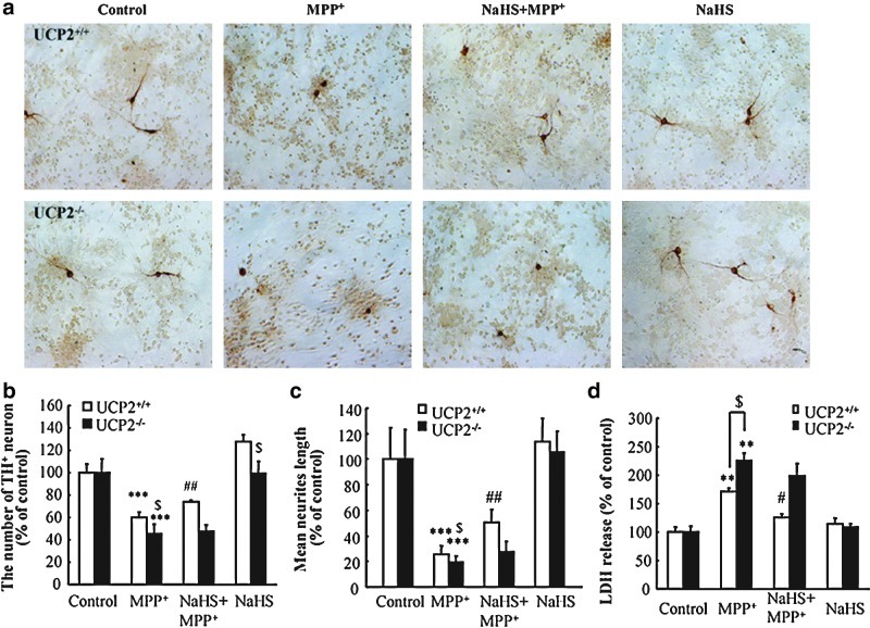
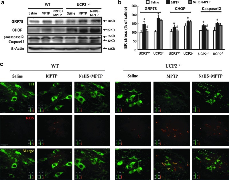
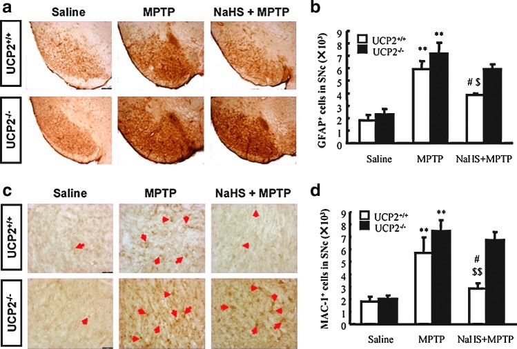
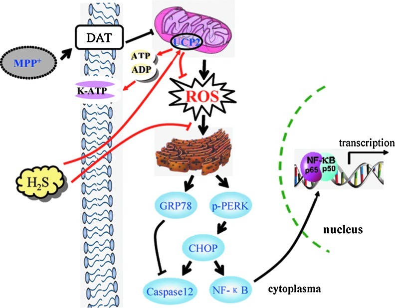
Results: Systemic administration of sodium hydrosulfide (NaHS) (an H(2)S donor, 5.6 mg/kg/day) for 7 days rescued MPTP-induced loss of dopaminergic (DA) neurons in substantia nigra compacta of both Kir6.2(+/+) and Kir6.2(-/-) mice. Consistently, NaHS (100 μM) protected primary mesencephalic neurons against 1-methyl-4-phenylpyridinium (MPP(+))-induced cytotoxicity in both genotypes. We further found that deficiency of mitochondrial uncoupling protein 2 (UCP2), which reduces reactive oxygen species (ROS) production and functions as upstream to the K-ATP channel in determining vulnerability of DA neurons, abolished the protective effects of H(2)S against either DA neuron degeneration in the PD mouse model or MPP(+)-induced injury in primary mesencephalic neurons. Rationally, UCP2 evokes mild uncoupling, which in turn diminishes ROS accumulation in DA neurons. Furthermore, H(2)S exerted neuroprotective effect via enhancing UCP2-mediated antioxidation and subsequently suppressing ROS-triggered endoplasmic reticulum stress as well as ultimately inhibiting caspase 12-induced neuronal apoptosis.

Innovation and conclusion: H(2)S protects DA neurons against degeneration in a UCP2 rather than Kir6.2/K-ATP channel-dependent mechanism, which will give us an insight into the potential of H(2)S in terms of opening up new therapeutic avenues for PD.


Figures

FIG. 1.
FIG. 1.
NaHS protected DA neurons against MPTP-induced degeneration in midbrain of wild type and Kir6.2−/− mice. Immunohistochemical staining of THir neurons in SNc of mice with ×40 magnifications (a). Stereological counts of THir cells in mouse SNpc (b) and VTA (c). Data are presented as the mean±S.E.M., n=5 for each group. NaHS had no significant effect on MPTP-reduced levels of DA (d), DOPAC (e), and HVA (f) in the striatum of both genotypic mice. Data expressed as means±S.E.M. from eight mice per group. *p<0.05, **p<0.01, and ***p<0.001 versus saline group in corresponding genotype; #p<0.05 and ##p<0.01 versus corresponding MPTP-treated groups. MPTP, 1-methyl-4-phenyl-1,2,3,6-tetrahydropyridine; DA, dopaminergic; TH, tyrosine-hydroxylase; TH+, tyrosine-hydroxylase positive; THir, tyrosine-hydroxylase immunoreactive; VTA, ventral tegmental area; DOPAC, dihydroxyphenylacetic acid; HVA, homovanillic acid; NaHS, sodium hydrosulfide; SNc, substantia nigra compacta; SNpc, substantia nigra pars compacta; S.E.M., standard error of mean. (To see this illustration in color the reader is referred to the web version of this article at www.liebertonline.com/ars).
FIG. 2.
FIG. 2.
NaHS protected against MPP+-induced cytotoxicity in primary cultured mesencephalic neurons from wild-type and Kir6.2−/− mice. (a) Typical photograph of immunocytochemical staining showing THir neurons at ×100 magnifications. (b, c) Effects of NaHS on the number of TH+ neurons (b) and LDH release (c) in MPP+-treated DA neurons. (d) Typical western blotting revealed that NaHS alleviated ERS and apoptosis in primary mesencephalic neurons isolated from wild-type and Kir6.2−/− mice. Statistical analysis of GRP78 (e), caspase 12 (f), and p65/H3 (g) protein levels in neurons. Data are presented as the mean±S.E.M. of four individual experiments. **p<0.01 and *p<0.05 versus control group; ##p<0.01 and #p<0.05 versus corresponding MPP+-treated groups; $p<0.05 versus Kir6.2−/− group. ERS, endoplasmic reticulum stress; GRP, glucose-regulated protein; LDH, lactate dehydrogenase; MPP+, 1-methyl-4-phenylpyridinium. (To see this illustration in color the reader is referred to the web version of this article at www.liebertonline.com/ars).
FIG. 3.
FIG. 3.
NaHS attenuated MPTP-triggered glial cell activation and proliferation in the SN of wild-type and Kir6.2−/− mice. (a) Microphotographs of GFAP-ir cells in the SN with ×40 magnification. (b) Stereological counts of GFAP-ir cells in mouse SN. (c) Microphotographs of Mac1-ir cells in the SN with ×200 magnification. (d) Stereological counts of Mac-1-ir cells in mouse SN. n=5. Data are presented as the mean±S.E.M., **p<0.01 and *p<0.05 versus saline group; #p<0.05 versus corresponding MPTP-treated groups; $p<0.05 versus corresponding MPTP-treated Kir6.2−/− groups. SN, substantia nigra; GFAP, Glial fibrillary acidic protein. Arrows indicate Mac-1+ cells. (To see this illustration in color the reader is referred to the web version of this article at www.liebertonline.com/ars).
FIG. 4.
FIG. 4.
UCP2 deficiency abolished the protective effect of NaHS on MPTP-injured TH+ neurons in SNc and VTA. (a) Microphotographs of THir neurons in SNpc of mice with ×40 magnification. Stereological counts of THir cells in SNpc (b) and VTA (c). Data are presented as the mean±S.E.M., n=5 for each group. *p<0.05 and **p<0.01 versus saline group in corresponding genotype; #p<0.05 versus corresponding MPTP-treated Ucp2+/+ groups; $p<0.05 versus corresponding Ucp2+/+ groups. UCP2, uncoupling protein 2. (To see this illustration in color the reader is referred to the web version of this article at www.liebertonline.com/ars).
FIG. 5.
FIG. 5.
UCP2 knockout abolished the protective effect of NaHS on MPP+-induced neurotoxicity in primary cultured mesencephalic neurons. THir neurons were stained by immunocytochemistry with ×200 magnifications (a). Effects of NaHS on MPP+-induced neurotoxicity in DA neurons as assessed by the number of TH+ neurons (b), mean neurites length per cell (c), and LDH release (d). Four independent experiments were performed in duplicate. Data were presented as the mean±S.E.M. ***p<0.001 and **p<0.01 versus control group; ##p<0.01 and #p<0.05 versus MPTP-treated Ucp2+/+ group; $p<0.05 Ucp.2+/+ group versus Ucp2−/− group. (To see this illustration in color the reader is referred to the web version of this article at www.liebertonline.com/ars).
FIG. 6.
FIG. 6.
UCP2 mediated the suppressive effect of NaHS on ROS-triggered ERS in the midbrain. Western blotting analysis of GRP78, CHOP, and caspase 12 protein levels in the midbrain of mice (a, b). UCP2 knockout abolished the inhibitory effect of NaHS on MPTP-induced ROS accumulation in SNc TH+ neurons (c). Photos were taken at ×600 magnification. Green color represents TH neuron and red color represents ROS. Scale bar=22 μm. Data are presented as the mean±S.E.M., n=4 for each group. *p<0.05 versus saline group; #p<0.05 versus corresponding MPTP-treated groups. ROS, reactive oxygen species; CHOP, C/EBP homologous protein. (To see this illustration in color the reader is referred to the web version of this article at www.liebertonline.com/ars).
FIG. 7.
FIG. 7.
UCP2 deletion abolished the inhibitory effect of NaHS on MPTP-triggered glial cell activation and proliferation. (a) Microphotographs of GFAP-ir cells in the SN with ×40 magnification. (b) Stereological counts of GFAP-ir cells in mouse SN. (c) Microphotographs of Mac1-ir cells in the SN with ×200 magnification. (d) Stereological counts of Mac-1-ir cells in mouse SN. n=4. Data are presented as the mean±S.E.M. **p<0.01 versus saline group; #p<0.05 versus corresponding MPTP-treated groups; $p<0.05 and $$p<0.01 versus corresponding Ucp2−/− groups. Arrows indicate Mac-1+ cells. Scale bar=100 μm. (To see this illustration in color the reader is referred to the web version of this article at www.liebertonline.com/ars).
FIG. 8.
FIG. 8.
Schematic model for the mechanism of H2S in protecting against MPP+-induced neuronal injury. MPP+ is selectively transported into DA neurons by DAT and subsequently inhibits the activity of mitochondrial complex I. Accumulated ROS evokes the upregulation of GRP78 and further triggers ERS. Consequently, p65 nuclear translocation-induced gene transcription or caspase 12-mediated apoptosis occurs in DA neurons. H2S-stimulated mitochondria uncoupling reduces the ratio of ATP/ADP, which in turn opens K-ATP channels. On the other hand, H2S enhances UCP2 function and attenuates ROS production, suppressing ERS and apoptosis induced by MPP+. DAT, dopamine transporter; K-ATP, adenosine triphosphate-sensitive potassium; H2S, hydrogen sulfide. (To see this illustration in color the reader is referred to the web version of this article at www.liebertonline.com/ars).

Similar articles

Cited by

References

    1. Abe K. Kimura H. The possible role of hydrogen sulfide as an endogenous neuromodulator. J Neurosci. 1996;16:1066–1071. - PMC - PubMed
    1. Bannenberg GL. Vieira HL. Therapeutic applications of the gaseous mediators carbon monoxide and hydrogen sulfide. Expert Opin Ther Pat. 2009;19:663–682. - PubMed
    1. Deutch AY. Winder DG. A channel to neurodegeneration. Nat Med. 2006;12:17–18. - PubMed
    1. Holtz WA. O'Malley KL. Parkinsonian mimetics induce aspects of unfolded protein response in death of dopaminergic neurons. J Biol Chem. 2003;278:19367–19377. - PubMed
    1. Hong Y. Fink BD. Dillon JS. Sivitz WI. Effects of adenoviral overexpression of uncoupling protein-2 and -3 on mitochondrial respiration in insulinoma cells. Endocrinology. 2001;142:249–256. - PubMed

Publication types

MeSH terms

LinkOut - more resources