

Current advances in detection and treatment of babesiosis
- PMID: 22360483
- PMCID: PMC3355466
- DOI: 10.2174/092986712799828355
Current advances in detection and treatment of babesiosis
Abstract
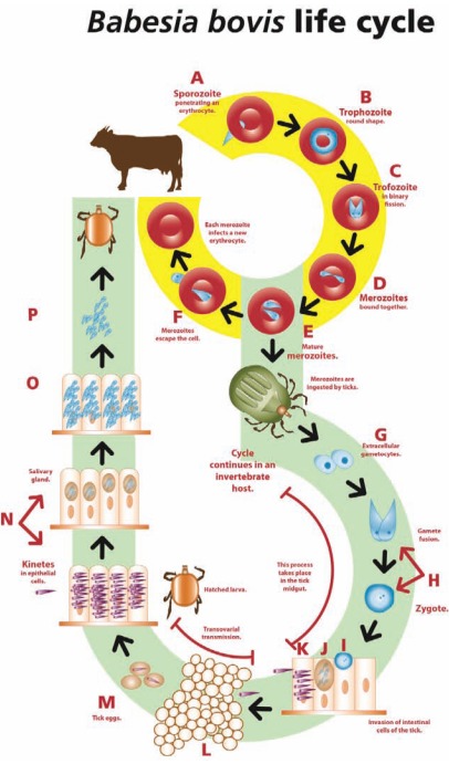
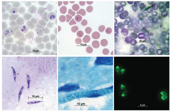
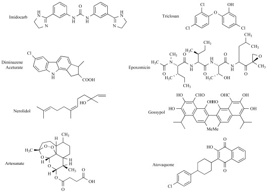
Babesiosis is a disease with a world-wide distribution affecting many species of mammals principally cattle and man. The major impact occurs in the cattle industry where bovine babesiosis has had a huge economic effect due to loss of meat and beef production of infected animals and death. Nowadays to those costs there must be added the high cost of tick control, disease detection, prevention and treatment. In almost a century and a quarter since the first report of the disease, the truth is: there is no a safe and efficient vaccine available, there are limited chemotherapeutic choices and few low-cost, reliable and fast detection methods. Detection and treatment of babesiosis are important tools to control babesiosis. Microscopy detection methods are still the cheapest and fastest methods used to identify Babesia parasites although their sensitivity and specificity are limited. Newer immunological methods are being developed and they offer faster, more sensitive and more specific options to conventional methods, although the direct immunological diagnoses of parasite antigens in host tissues are still missing. Detection methods based on nucleic acid identification and their amplification are the most sensitive and reliable techniques available today; importantly, most of those methodologies were developed before the genomics and bioinformatics era, which leaves ample room for optimization. For years, babesiosis treatment has been based on the use of very few drugs like imidocarb or diminazene aceturate. Recently, several pharmacological compounds were developed and evaluated, offering new options to control the disease. With the complete sequence of the Babesia bovis genome and the B. bigemina genome project in progress, the post-genomic era brings a new light on the development of diagnosis methods and new chemotherapy targets. In this review, we will present the current advances in detection and treatment of babesiosis in cattle and other animals, with additional reference to several apicomplexan parasites.
Figures
References
-
- Ristic M. In: Diseases of Cattle in the Tropics. Firts Edition ed. Ristic M, McIntyre I, editors. Vol. 6. The Hague: Martinus Nijhoff Publishers; 1981. pp. 443–468.
-
- Bock R, Jackson L, de Vos A, Jorgensen W. Babesiosis of cattle. Parasitology. 2004;(129 Suppl):S247–269. - PubMed
-
- Brayton KA, Lau AO, Herndon DR, Hannick L, Kappmeyer LS, Berens SJ, Bidwell SL, Brown WC, Crabtree J, Fadrosh D, Feldblum T, Forberger HA, Haas BJ, Howell JM, Khouri H, Koo H, Mann DJ, Norimine J, Paulsen IT, Radune D, Ren Q, Smith RK, Jr, Suarez CE, White O, Wortman JR, Knowles DP, Jr, McElwain TF, Nene VM. Genome sequence of Babesia bovis and comparative analysis of apicomplexan hemoprotozoa. PLoS pathogens. 2007;3(10):1401–1413. - PMC - PubMed
-
- Babes V. Sur l' hémoglobinurie bactérienne du boeuf. C. R. Acad. Sci. 1888;107:692–694.
Publication types
MeSH terms
Substances
LinkOut - more resources
Full Text Sources