. 2012 Feb 23:10:15.
doi: 10.1186/1477-7827-10-15.

Down-regulation of the transcription factor snail in the placentas of patients with preeclampsia and in a rat model of preeclampsia

Affiliations

Down-regulation of the transcription factor snail in the placentas of patients with preeclampsia and in a rat model of preeclampsia

Larisa Fedorova et al. Reprod Biol Endocrinol. .

Abstract

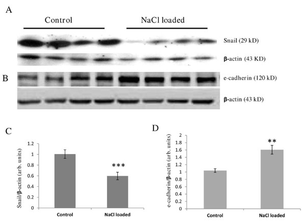
Background: Placental malfunction in preeclampsia is believed to be a consequence of aberrant differentiation of trophoblast lineages and changes in utero-placental oxygenation. The transcription factor Snail, a master regulator molecule of epithelial-mesenchymal transition in embryonic development and in cancer, is shown to be involved in trophoblast differentiation as well. Moreover, Snail can be controlled by oxidative stress and hypoxia. Therefore, we examined the expression of Snail and its downstream target, e-cadherin, in human normal term, preterm and preeclamptic placentas, and in pregnant rats that developed preeclampsia-like symptoms in the response to a 20-fold increase in sodium intake.

Methods: Western blotting analysis was used for comparative expression of Snail and e- cadherin in total protein extracts. Placental cells expressing Snail and e-cadherin were identified by immunohistochemical double-labeling technique.

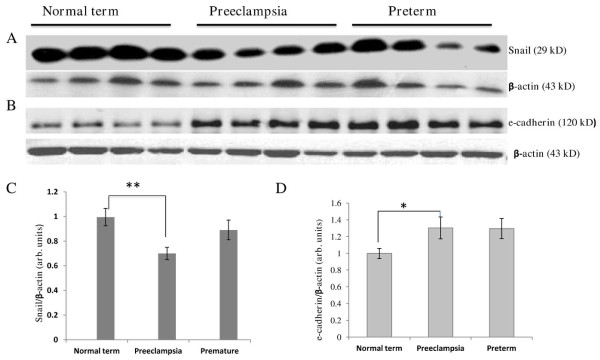
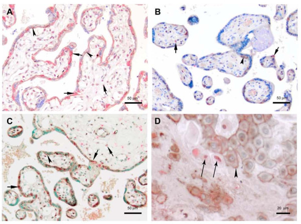
Results: The levels of Snail protein were decreased in human preeclamptic placentas by 30% (p < 0.01) compared to normal term, and in the rat model by 40% (p < 0.001) compared to control placentas. In preterm placentas, the levels of Snail expression varied, yet there was a strong trend toward statistical significance between preterm and preeclamptic placentas. In humans, e-cadherin protein level was 30% higher in preeclamptic (p < 0.05) placentas and similarly, but not significantly (p = 0.1), high in the preterm placentas compared to normal term. In the rat model of preeclampsia, e-cadherin was increased by 60% (p < 0.01). Immunohistochemical examination of human placentas demonstrated Snail-positive staining in the nuclei of the villous trophoblasts and mesenchymal cells and in the invasive trophoblasts of the decidua. In the rat placenta, the majority of Snail positive cells were spongiotrophoblasts of the junctional zone, while in the labyrinth, Snail-positive sinusoidal giant trophoblasts cells were found in some focal areas located close to the junctional zone.

Conclusion: We demonstrated that human preeclampsia and the salt-induced rat model of preeclampsia are associated with the reduced levels of Snail protein in placenta. Down-regulation of the transcription factor Snail in placental progenitor cell lineages, either by intrinsic defects and/or by extrinsic and maternal factors, may affect normal placenta development and function and thus contribute to the pathology of preeclampsia.


Figures

Figure 1
Figure 1
Western blotting analysis of Snail and e-cadherin expression in the total protein extracts from human placentas. A representative immunoblot is shown (Snail) from at least four separate runs of each sample (A), and graph (C) demonstrating densitometry analysis of Snail protein levels in normal term, preeclamptic, and preterm placentas (n = 36, 32, 30 respectively). For Snail, the ANOVA test revealed a significant difference among the three groups (p value = 0.0100). However, the Tukey HSD post-hoc test only indicated a statistically significant difference when comparing the normal term group and the preeclampsia group (p value = 0.0085). The difference between the preterm and the preeclamptic samples was not significant (p = 0.0941). Representative immunoblot (e-cadherin) from three separate gels (B), and graph showing densitometry analysis of e-cadherin levels in the three groups studied (D) (n = 27, 24 and 15 for normal term, preeclamptic, and preterm placentas respectively). For e-cadherin, the ANOVA test also revealed a significant difference among the three groups (p value = 0.0265). Again, the Tukey HSD post-hoc test only indicated a statistically significant difference when comparing the normal term group and the preeclampsia group (p value = 0.0397). The difference between the preterm and normal term group was not statistically significant (p = 0.1). * p < 0.05; ** p < 0.01
Figure 2
Figure 2
Immunohistochemical analysis of Snail and e-cadherin proteins expression in human normal term, preeclamptic and preterm placentas. E-cadherin (stained brown) is present in the basement membranes of the villi and, to a lesser extent, in the cytoplasm of VCT where it forms a connection between neighboring VCTs, and between VCT and overlying SynTB (arrowheads). Snail (stained red) is localized mostly in the nuclei of VCT and mesenchymal cells (short arrows). A normal placenta (A) and a preeclamptic placenta (B) are shown. In preeclampsia, there is a significant reduction in Snail expression. The premature placenta (C) shows a similar staining pattern to the term placenta. The maternal-fetal interface of a preterm placenta (D) reveals expression of Snail in EVT (long arrows), which lack e-cadherin on their membranes. Note that EVTs which express e-cadherin are Snail negative. The loss of e-cadherin by EVT is believed necessary for proper invasion and implantation
Figure 3
Figure 3
Western blotting analysis of Snail and e-cadherin expression in the total protein extracts from rat placentas. Representative immunoblot from 3 independent runs for each sample (Snail) (A), and graph demonstrating densitometry analysis of Snail protein levels in the placentas of control pregnant rats and rats loaded with NaCl (n ≥ 15) (C). Representative immunoblot from 2 independent runs for each sample (e-cadherin) (B), and graph showing densitometry analysis of e-cadherin levels in the two groups studied (n ≥ 10) (D).**p < 0.01, *** p < 0.001
Figure 4
Figure 4
Immunohistochemistry of Snail and e-cadherin expression in normal definitive rat placenta. The photomicrographs show that Snail (stained red) is present in the nuclei of spongiotrophoblast cells of the junctional zone. E-cadherin (stained brown) is localized to the SynTB of the labyrinth (A). Snail positivity is also found in mononuclear sinusoidal TGC in some sinusoids positioned close to the junctional zone (arrow) (B)

Similar articles

Cited by

References

    1. Steegers EA, von Dadelszen P, Duvekot JJ, Pijnenborg R. Pre-eclampsia. Lancet. 2010;376(9741):631–644. doi: 10.1016/S0140-6736(10)60279-6. - DOI - PubMed
    1. Benirschke K, Kaufmann P, Baergen R. Pathology of the human placenta. 5. New York: Springer; 2006.
    1. Huppertz B. Placental origins of preeclampsia: challenging the current hypothesis. Hypertension. 2008;51(4):970–975. doi: 10.1161/HYPERTENSIONAHA.107.107607. - DOI - PubMed
    1. Burton GJ, Jones CJ. Syncytial knots, sprouts, apoptosis, and trophoblast deportation from the human placenta. Taiwan J Obstet Gynecol. 2009;48(1):28–37. doi: 10.1016/S1028-4559(09)60032-2. - DOI - PubMed
    1. Huppertz B. IFPA Award in Placentology Lecture: biology of the placental syncytiotrophoblast-myths and facts. Placenta. 2010;31(Suppl):S75–S81. - PubMed

Publication types