. 2012 Mar 23;36(3):464-76.
doi: 10.1016/j.immuni.2012.02.001. Epub 2012 Feb 21.

An NLRP7-containing inflammasome mediates recognition of microbial lipopeptides in human macrophages

Affiliations

An NLRP7-containing inflammasome mediates recognition of microbial lipopeptides in human macrophages

Sonal Khare et al. Immunity. .

Abstract

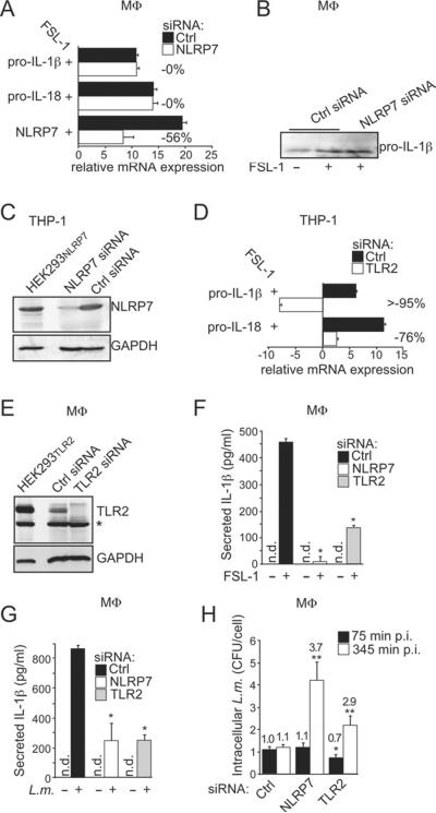
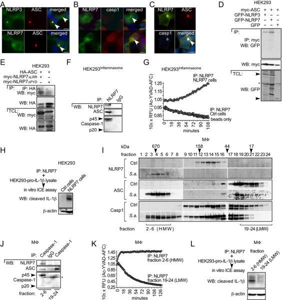
Cytosolic pathogen- and damage-associated molecular patterns are sensed by pattern recognition receptors, including members of the nucleotide-binding domain and leucine-rich repeat-containing gene family (NLR), which cause inflammasome assembly and caspase-1 activation to promote maturation and release of the inflammatory cytokines interleukin-1β (IL-1β) and IL-18 and induction of pyroptosis. However, the contribution of most of the NLRs to innate immunity, host defense, and inflammasome activation and their specific agonists are still unknown. Here we describe identification and characterization of an NLRP7 inflammasome in human macrophages, which is induced in response to microbial acylated lipopeptides. Activation of NLRP7 promoted ASC-dependent caspase-1 activation, IL-1β and IL-18 maturation, and restriction of intracellular bacterial replication, but not caspase-1-independent secretion of the proinflammatory cytokines IL-6 and tumor necrosis factor-α. Our study therefore increases our currently limited understanding of NLR activation, inflammasome assembly, and maturation of IL-1β and IL-18 in human macrophages.


Figures

Figure 1
Figure 1. MP causes ASC translocation form the nucleus to the cytosol and secretion of IL-1β from MΦ
(A,B) (A) THP-1 cells were treated with culture SN from HEK293 cells either negative (Ctrl, panel 1) or positive for MP for 30 min (panel 2) or for 16 hrs (panel 3), treated with HKAL (panel 4), HKLP (panel 5) or HKSA (panel 6) for 6 hrs or (B) MΦ were either mock treated (upper panel) or treated with HKAL (lower panel) for 6 hrs and immunostained for ASC, DNA and actin. The scale bar is 20 μm. (C) Mock and MP infected THP-1 cells were separated into nuclear (Nuc) and cytosolic (Cyt) fractions and analyzed by Western blot (WB). (D,E) MΦ were treated with vehicle Ctrl or HKAL for 16 hrs and (D) SN were analyzed for IL-1β (n=3 ±SD) or (E) TCL and concentrated SN were analyzed by WB. (FH) THP-1 cells were treated with (F) vehicle Ctrl or HK bacteria for 16 hrs or (G) with Ctrl or MP positive culture SN for 24 hrs. SN were analyzed for IL-1β as above or (H) analyzed for the presence of MP (OD600 ≥ 1.0 indicates the presence of MP); n=3 ±SD; *p≤0.05.
Figure 2
Figure 2. ASC and NLRP7 are required for HKAL and acLP-induced IL-1β secretion
(A,B) THP-1shLuc, THP-1shCtr and THP-1shASC cells were treated with (A) vehicle Ctrl or HK bacteria (B) vehicle Ctrl, HKAL or acLP for 16 hrs and SN were analyzed for IL-1β (n=3 ±SD). TCL were analyzed by WB. (CF) THP-1 cells were transfected with (C,D) pooled or (E, F) individual non-targeting siRNAs (Ctrl) and siRNAs targeting NLRPs, as indicated and treated with HKAL for 16 hrs; (C,E) mRNA expression of NLRPs was analyzed by RT-PCR and β-actin control; (D,F) SN were analyzed for IL-1β as above. n=3, ±SD (G,H) MΦ were transfected with Ctrl or NLRP-specific siRNAs, treated with HKAL or FSL-1 for 16 hrs; (G) IL-1β in SN was determined (n=3 ±SD) and (H) Silencing of NLRP expression was confirmed by RT-PCR. (IK) (I) THP-1 cells or (J,K) MΦ were transfected using Ctrl or NLRP7 siRNAs, mock- or acLP-treated, transfected with dA:dT or primed with ultrapure LPS for 6 hrs and infected with adenovirus (AdV) for 16 hrs, as indicated and (I,J) analyzed for IL-1β and (K) IL-18 release as above. n=3 ±SD; *p≤0.05
Figure 3
Figure 3. NLRP3 and NLRP7 recognize intracellular bacteria and restrict bacterial replication
(AD) MΦ were transfected using Ctrl and NLRP siRNAs, mock treated or treated with HK bacteria or FSL-1 for 16 hrs, as indicated and (A,B,D) analyzed for IL-1β (n=3 ±SD, *p≤0.05) or (C) analyzed by RT-PCR. (EH) MΦ were transfected using Ctrl, NLRP3 or NLRP7 siRNAs and left uninfected or were infected with (E,F) S. aureus (S.a) or (G,H) L. monocytogenes (L.m) and analyzed for (E,G) IL-1β 345 min p.i. (n=3 ±SD) or (F,H) were lysed at 75 and 345 min p.i. and intracellular colony forming units (CFU) were determined. Results from a RIP representative experiment are presented as CFU/cell and the fold increase compared to control siRNA transfected cells at 75 minutes p.i. is indicated. (I) THP-1 cells were transfected with siRNAs as indicated and kept uninfected or infected with S.a. as above. SN were analyzed at the indicated times for released LDH and presented as % cytotoxicit (n=3 ±SD; *p≤0.05); n.d.: not detectable.
Figure 4
Figure 4. NLRP7 mediates acLP-induced IL-1β release through caspase-1 activation
(A) MΦ were pretreated with vehicle Ctrl (DMSO), zYVAD-fmk or zLEHD-fmk for 1 hr, treated with vehicle Ctrl, HKAL or FSL-1 for 16 hrs and analyzed for IL-1β release (n=3 ±SD); *p≤0.05. (B,C) MΦ were not transfected (none), transfected with Ctrl or NLRP7 siRNAs. (B) Mock or cycloheximide (CHX)-treated (to prevent re-synthesis of pro-caspase-1) and FSL-1 or cLPS activated as indicated or (C) FSL-1-activated for 6 hrs and (B) TCL and (C) SN were analyzed by WB. (D,E) (D) MΦ or (E) THP-1 cells were transfected with Ctrl or NLRP7 siRNAs and activated with FSL-1 or cLPS for 16 hrs as indicated and SN were analyzed for (D) IL-6 and (E) TNFα by ELISA (n=3 ±SD). Cells were pre-treated for 1 hr with anakinra to prevent autocrine IL-1β signaling; n.d.: not detectable. (F) MΦ (upper panel) and THP-1 cells (lower panel) were transfected as indicated, activated with FSL-1, cLPS or TNF-α for the indicated times and TCL were analyzed by WB.
Figure 5
Figure 5. NLRP7 and TLR2 are required for acLP-induced IL-1β release
(A) MΦ were transfected with Ctrl or NLRP7 siRNAs, activated with FSL-1 for 16 hrs and analyzed by qPCR (relative expression compared to βactin, n=3 ±SD). Silencing efficiency is indicated in % compared to control siRNA. (B) MΦ were transfected with Ctrl or NLRP7 siRNAs, activated with FSL-1 for 16 hrs in the presence of zYVAD-fmk to prevent maturation of IL-1β and TCL were analyzed for pro-IL-1β by WB. (C) THP-1 cells were transfected with Ctrl or NLRP7 siRNAs and analyzed by WB, using NLRP7-transfected HEK293NLRP7 cells as control. (D) THP-1 cells were transfected with Ctrl or TLR2 siRNAs, treated with FSL-1 for 16 hrs and analyzed by qPCR for IL-1β and IL-18 mRNA, as described above. n=3 ±SD. (E,F) MΦ were transfected with Ctrl, NLRP7 or TLR2 siRNAs, mock treated or treated with FSL-1 for 16 hrs and (E) TCL were analyzed by WB using TLR2 transfected HEK293TLR2 cells as control. *cross-reactive protein; and (F) analyzed for IL-1β release (n=3 ±SD). (G,H) MΦ were transfected using Ctrl, NLRP7 or TLR2 siRNAs and either left uninfected or were infected with L. monocytogenes (L.m.) and (G) were analyzed for IL-1β release 345 min p.i., or (H) were lysed at 75 and 345 min p.i. and CFU were determined. Results are presented as CFU/cell and the fold increase compared to Ctrl siRNA transfected cells at 75 min p.i. is indicated (n=3 ±SD); n.d.: not detectable; *p≤0.05
Figure 6
Figure 6. Activation of NLRP7 causes the formation of a high-molecular weight inflammasome in MΦ
(AC) Immunofluorescence staining of HEK293 cells transiently transfected with (A) ASC and NLRP3 (upper panel), ASC and NLRP7 (lower panel), (B) NLRP7 and pro-caspase-1 (upper panel shows a cell expressing only NLRP7; lower panel shows cells expressing NLRP7 and caspase-1) or (C) NLRP7, ASC and pro-caspase-1. NLR-containing aggregates are marked by arrowheads. The scale bar is 20 μm. (D) HEK293ASC cells were transfected with GFP, GFP-NLRP3 or GFP-NLRP7 as indicated and TCL were used for IP with immobilized myc antibodies and WB as indicated. (E) HEK293 cells were transfected with HA-ASC, myc-NLRP7ΔLRR or myc-NLRP7ΔPYD, as indicated and TCL were used for IP with immobilized HA antibodies and WB as indicated. (FH) Stable HEK293 inflammasome reconstituted cells (HEK293Inflammasome) were transfected with control or NLRP7 as indicated, and TCL were used for IP with immobilized NLRP7 antibodies. Immune complexes were analyzed (F) by WB as indicated; (G) equilibrated in caspase-1 assay buffer and subjected to in vitro caspase-1 activity assay or (H) for ICE activity incubated with a TCL isolated from HEK293 cells transfected with pro-IL-1β and analyzed by WB for mature IL-1β. (IL) Ctrl or S.a.-infected MΦ were fractionated by SEC and pooled fractions were (I) TCA precipitated and analyzed by WB as indicated; (J) Caspase-1 was immunoprecipitated from fractions 2–6 and 19–24 of S.a.-infected MΦ using IgG as control and analyzed by WB as indicated; (K,L) NLRP7 was immunoprecipitated from the same fractions and (K) assayed for caspase-1 activity assay (activity purified from fractions 2–6 saturated the assay) or (L) assayed for ICE activity as above. *cross-reactive protein.
Figure 7
Figure 7. Reconstituted NLRP7 inflammasomes are sufficient to respond to HKAL and acLP
(A)HEK293 cells were transiently transfected as indicated and release of IL-1β was determined in SN. Results are presented as fold increase compared to cells transfected with pro-IL-1β and pro-caspase-1; (n=3 ±SD); *p≤0.05 compared to cells not transfected with NLRPs. (B) HEK293 cells were transfected as above with suboptimal concentrations of NLRP7, followed by a 2nd transfection after 24 hrs with transfection reagent alone (Ctrl), HKAL (8×10⁵ cfu) or FSL-1 (0.5 μg). Secreted IL-1β is presented as fold increase compared to control transfected cells. (n=3 ±SD); *p≤0.05 compared to cells lacking NLRP7; **p≤0.05 compared to the 2nd round of mock-transfected cells. (C) HEK293 cells were transfected as above with suboptimal concentrations of NLRP7, NLRP3, Nod1 or AFAP1, followed by FSL-1 transfection. Secreted IL-1β is presented as fold increase compared to Ctrl transfected cells; (n=3 ±SD); *p≤0.05 compared to cells lacking NLRs; **p≤0.05 compared to the 2nd round of mock-transfected cells. Expression of NLRP7 (118 kDa), NLRP3 (118 kDa), Nod1 (107 kDa), and AFAP1 (110 kDa) in the inflammasome reconstitution assay was verified by WB. (D) HEK293 cells were transfected and analyzed for IL-1β secretion as above, but with NLRP7 and NLRP7ΔLRR; (n=3 ±SD); *p≤0.05 compared to the 2nd round of mock-transfected cells. (E) HEK293 cells were transfected with NLRP7, NLRP7R693W, NLRP7R693P or NLRP7D657V, followed by FSL-1 transfection as above and secreted IL-1β is presented as fold increase compared to cells transfected with pro-IL-1β and pro-caspase-1; (n=3 ±SD); *p≤0.05 compared to cells not transfected with FSL-1; **p≤0.05 compared to cells transfected with NLRP7; ***p≤0.05 compared to cells transfected with NLRP7 and FSL-1. Expression of NLRP7 was verified by WB. (F) MΦ were transfected using Ctrl, NLRP3 or NLRP7 siRNAs, primed with ultra pure LPS for 4 hrs and either left untreated or lysosomal damage was inflicted with Leu-Leu-OMe for 16 hrs and analyzed for IL-1β release (n=6 ±SD). (G,H) THP-1 cells were pretreated with either medium containing 130 mM KCl (KCL) or a cathepsin B inhibitor (CA-074-Me), mock-treated or treated with MSU or (G) MSU or (H) FSL-1 for 8 hrs and secreted IL-1β was determined; (n=3 ±SD); ***p≤0.05.

Comment in

Similar articles

Cited by

References

    1. Alexopoulou L, Thomas V, Schnare M, Lobet Y, Anguita J, Schoen RT, Medzhitov R, Fikrig E, Flavell RA. Hyporesponsiveness to vaccination with Borrelia burgdorferi OspA in humans and in TLR1- and TLR2-deficient mice. Nat Med. 2002;8:878–884. - PubMed
    1. Aliprantis AO, Yang RB, Mark MR, Suggett S, Devaux B, Radolf JD, Klimpel GR, Godowski P, Zychlinsky A. Cell activation and apoptosis by bacterial lipoproteins through toll-like receptor-2. Science. 1999;285:736–739. - PubMed
    1. Bernthal NM, Pribaz JR, Stavrakis AI, Billi F, Cho JS, Ramos RI, Francis KP, Iwakura Y, Miller LS. Protective role of IL-1β against post-arthroplasty Staphylococcus aureus infection. J Orthopaed Res. 2011;29:1621–1626. - PMC - PubMed
    1. Boyden ED, Dietrich WF. Nalp1b controls mouse macrophage susceptibility to anthrax lethal toxin. Nat Genet. 2006;38:240–244. - PubMed
    1. Broz P, von Moltke J, Jones JW, Vance RE, Monack DM. Differential Requirement for Caspase-1 Autoproteolysis in Pathogen-Induced Cell Death and Cytokine Processing. Cell Host & Microbe. 2010;8:471–483. - PMC - PubMed

Publication types

MeSH terms