. 2012 May;235(1):246-55.
doi: 10.1016/j.expneurol.2012.02.002. Epub 2012 Feb 14.

Decreased expression of the glial water channel aquaporin-4 in the intrahippocampal kainic acid model of epileptogenesis

Affiliations

Decreased expression of the glial water channel aquaporin-4 in the intrahippocampal kainic acid model of epileptogenesis

Darrin J Lee et al. Exp Neurol. 2012 May.

Abstract

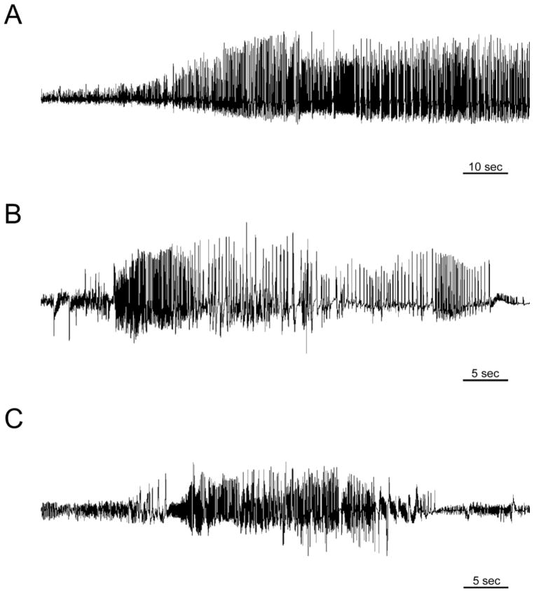
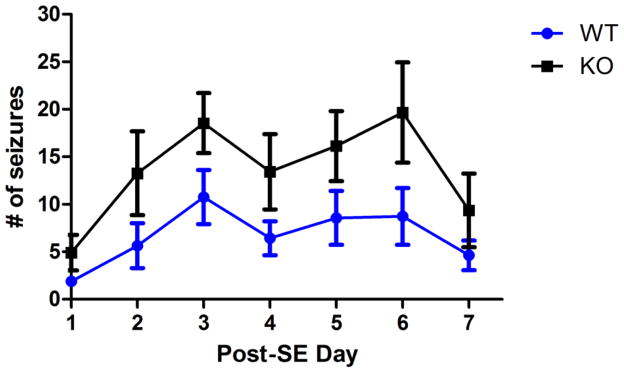
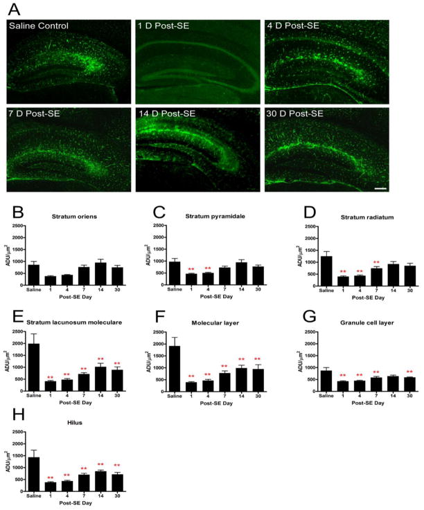
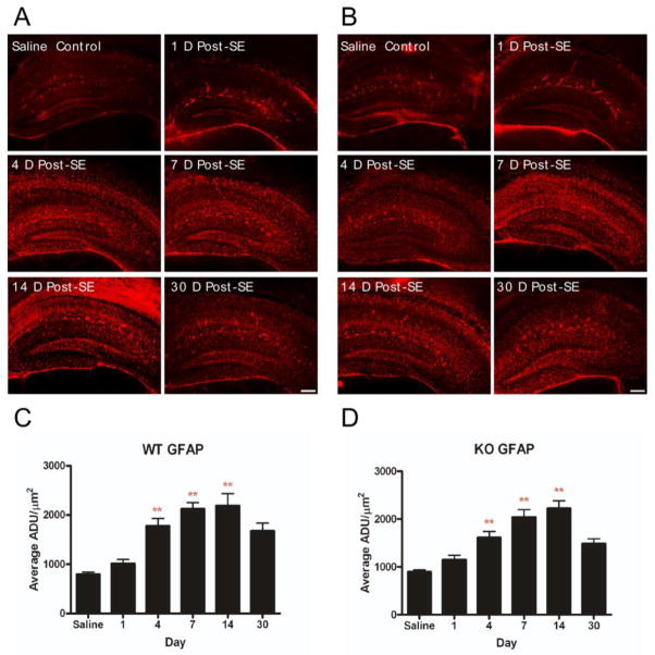
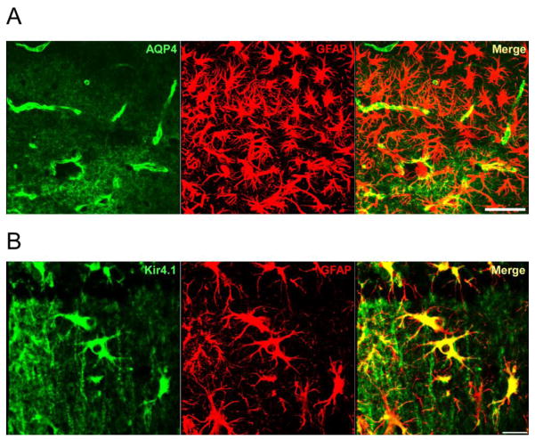
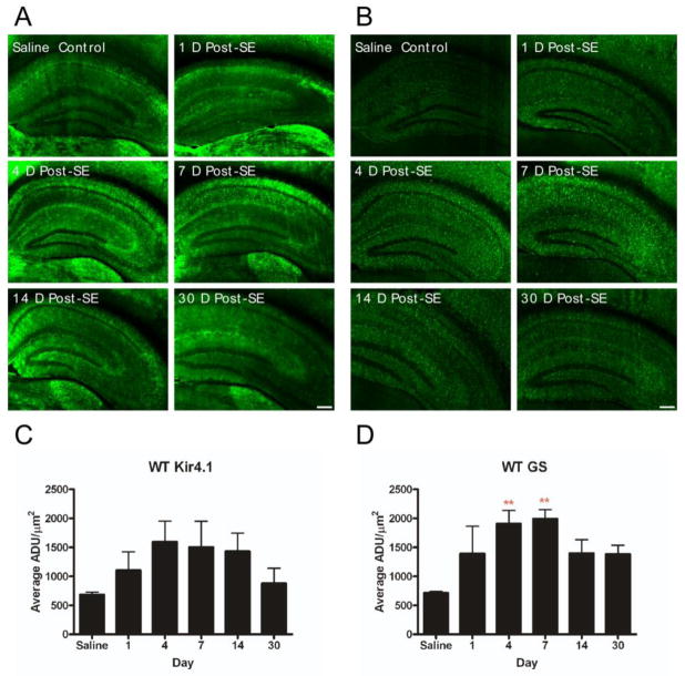
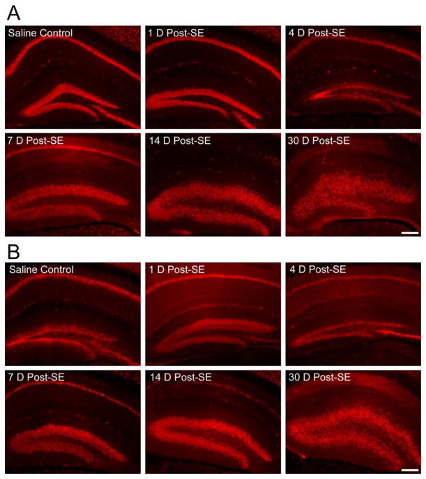
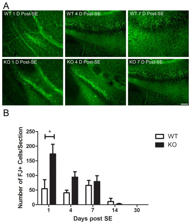
Recent evidence suggests that astrocytes may be a potential new target for the treatment of epilepsy. The glial water channel aquaporin-4 (AQP4) is expressed in astrocytes, and along with the inwardly-rectifying K(+) channel K(ir)4.1 is thought to underlie the reuptake of H(2)O and K(+) into glial cells during neural activity. Previous studies have demonstrated increased seizure duration and slowed potassium kinetics in AQP4(-/-) mice, and redistribution of AQP4 in hippocampal specimens from patients with chronic epilepsy. However, the regulation and role of AQP4 during epileptogenesis remain to be defined. In this study, we examined the expression of AQP4 and other glial molecules (GFAP, K(ir)4.1, glutamine synthetase) in the intrahippocampal kainic acid (KA) model of epilepsy and compared behavioral and histologic outcomes in wild-type mice vs. AQP4(-/-) mice. Marked and prolonged reduction in AQP4 immunoreactivity on both astrocytic fine processes and endfeet was observed following KA status epilepticus in multiple hippocampal layers. In addition, AQP4(-/-) mice had more spontaneous recurrent seizures than wild-type mice during the first week after KA SE as assessed by chronic video-EEG monitoring and blinded EEG analysis. While both genotypes exhibited similar reactive astrocytic changes, granule cell dispersion and CA1 pyramidal neuron loss, there were an increased number of fluorojade-positive cells early after KA SE in AQP4(-/-) mice. These results indicate a marked reduction of AQP4 following KA SE and suggest that dysregulation of water and potassium homeostasis occurs during early epileptogenesis. Restoration of astrocytic water and ion homeostasis may represent a novel therapeutic strategy.


Figures

Figure 1
Figure 1
Intrahippocampal kainic acid model. A. EEG recording during status epilepticus. Within the first 30 minutes after kainic acid injection, the mouse develops electrographic and behavioral status epilepticus. B. Example of EEG recording of a spontaneous seizure in an AQP4+/+ mouse. C. Example of EEG recording of a spontaneous seizure in an AQP4−/− mice. Within 2–7 days post-injection, both AQP4+/+ and AQP4−/− mice which underwent status epilepticus developed spontaneous seizures.
Figure 2
Figure 2
Increased seizure frequency in AQP4−/− mice. Both AQP4+/+ and AQP4−/− mice developed spontaneous seizures after undergoing status epilepticus; however, AQP4−/− mice exhibited more seizures during the epileptogenic period (post-status epilepticus days 1–7, 2-way RM ANOVA, p<0.05).
Figure 3
Figure 3
AQP4 immunoreactivity following kainic acid status epilepticus. Significant reduction in hippocampal AQP4 immunoreactivity was observed with delayed partial recovery. A. 4x images at indicated time points after SE. Scale bar = 200 μm. Laminar-specific analysis of AQP4 immunoreactivity after SE demonstrates decreased AQP4 immunoreactivity detected in various layers of the hippocampus throughout the study period (B-H). The initial decrease in AQP4 immunoreactivity is followed by a gradual increase. Persistent downregulation was observed in the SLM, ML, granule cell layer and hilus. ADU= arbitrary density units. **, p<0.01 compared to saline control.
Figure 4
Figure 4
Immunohistochemical staining of GFAP. Increased GFAP immunoreactivity and astrocyte ramification occurs following SE in both AQP4+/+ (A, C) and AQP4−/− (B, D) mice. Scale bar = 200 μm.
Figure 5
Figure 5
Comparison of AQP4 and Kir4.1 labeling of reactive astrocytes. A. Confocal 40x image in CA1 stratum lacunosum moleculare 7 days post-SE. Note marked reactive phenotype of GFAP-positive astrocytes (center) but lack of strong AQP4 immunoreactivity. Scale bar = 50μm. B. Confocal 63x image of reactive astrocytes in CA1 stratum radiatum 14 days post-SE. Note strong Kir4.1 immunoreactivity on reactive astrocytes. Scale bar = 20μm.
Figure 6
Figure 6
Kir4.1 and glutamine synthetase immunoreactivity. A, C. No significant overall change in Kir4.1 immunoreactivity was observed post-SE. However, focal increase in Kir4.1 immunoreactivity was detected within the ramified astrocytes of the stratum radiatum and stratum lacunosum moleculare during post-SE days 4 and 7 (Supplemental Figure 2). B, D. Glutamine synthetase immunoreactivity was transiently elevated at post-SE days 4 and 7. Scale bar = 200μm.
Figure 7
Figure 7
Granule cell dispersion and CA1 pyramidal cell loss following intrahippocampal KA administration. After KA-induced SE, granule cell dispersion was noted in both AQP4+/+ (A) and AQP4−/− (B) mice as early as post-SE day 7. Granule cell dispersion was observed at post-SE day 14 and 30, and is accompanied by loss of NeuN immunoreactivity in the CA1 pyramidal cell layer. Scale bar = 200μm.
Figure 8
Figure 8
Fluoro-jade-B histochemistry following SE. A. Representative examples of FJ-B-positive cells in the CA3 region and hilus of the dentate gyrus in the hippocampus ipsilateral to the injection in AQP4+/+ and AQP4−/− mice. Scale bar = 100μm. B. There were significantly more FJ-B-positive cells within AQP4−/− hippocampi relative to AQP4+/+ hippocampi on post-SE day 1 (2-way ANOVA with post-hoc Bonferroni test, P<0.001).

Similar articles

Cited by

References

    1. Amiry-Moghaddam M, Williamson A, Palomba M, Eid T, de Lanerolle NC, Nagelhus EA, Adams ME, Froehner SC, Agre P, Ottersen OP. Delayed K+ clearance associated with aquaporin-4 mislocalization: phenotypic defects in brains of alpha-syntrophin-null mice. Proceedings of the National Academy of Sciences. 2003;100:13615–13620. - PMC - PubMed
    1. Andrew RD, Fagan M, Ballyk BA, Rosen AS. Seizure susceptibility and the osmotic state. Brain Res. 1989;498:175–180. - PubMed
    1. Arabadzisz D, Antal K, Parpan F, Emri Z, Fritschy JM. Epileptogenesis and chronic seizures in a mouse model of temporal lobe epilepsy are associated with distinct EEG patterns and selective neurochemical alterations in the contralateral hippocampus. Experimental Neurology. 2005;194:76–90. - PubMed
    1. Ballanyi K, Grafe P, ten Bruggencate G. Ion activities and potassium uptake mechanisms of glial cells in guinea-pig olfactory cortex slices. J Physiol. 1987;382:159–174. - PMC - PubMed
    1. Binder DK, Oshio K, Ma T, Verkman AS, Manley GT. Increased seizure threshold in mice lacking aquaporin-4 water channels. Neuroreport. 2004;15:259–262. - PubMed

Publication types