. 2012 Jul;236(1):6-18.
doi: 10.1016/j.expneurol.2012.02.006. Epub 2012 Feb 14.

The lateral thoracic nerve and the cutaneous maximus muscle--a novel in vivo model system for nerve degeneration and regeneration studies

Affiliations

The lateral thoracic nerve and the cutaneous maximus muscle--a novel in vivo model system for nerve degeneration and regeneration studies

Baohan Pan et al. Exp Neurol. 2012 Jul.

Abstract

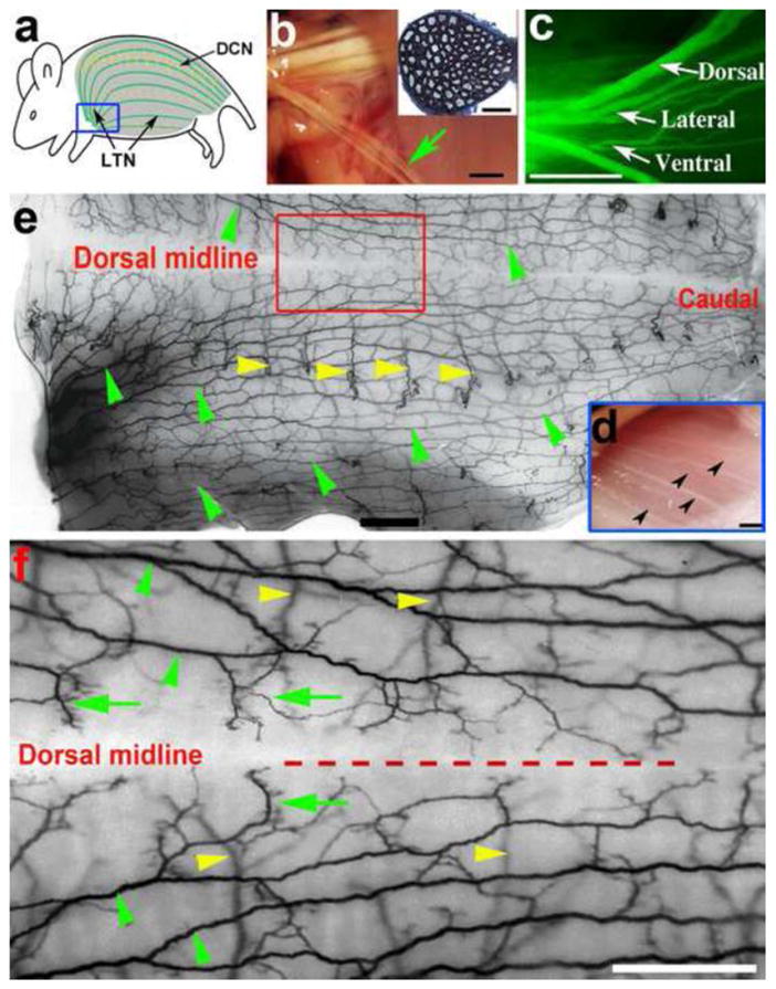
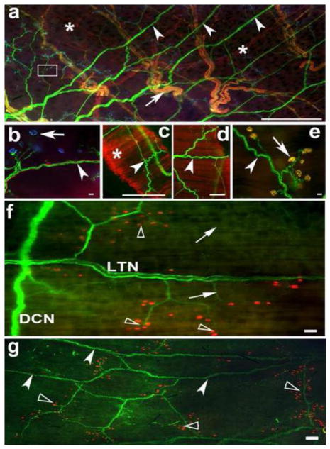
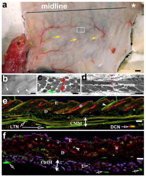
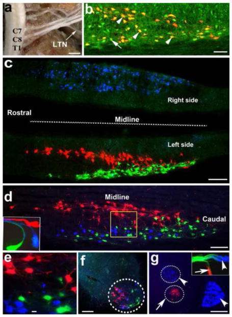
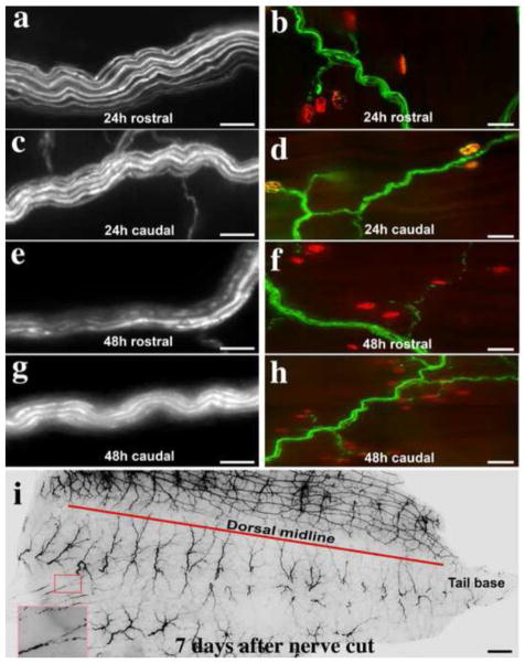
We report a novel in vivo mouse model system to study regeneration of injured motor nerve and spatiotemporal pattern of denervation in experimental nerve diseases. The lateral thoracic nerve (LTN), as a pure motor nerve, innervates the cutaneous maximus muscle (CMM) by some of the shortest and the longest motor nerve fibers in the mouse body. Its branches and nerve terminals can be imaged in whole mount preparations. Here we describe the branching pattern of the LTN and its innervation of the CMM, and characterize degeneration and regeneration over time after a LTN crush by morphological and electrophysiological analyses. We demonstrate the utility of this model in a well-established neurotoxicity paradigm and in a genetic disease model of the peripheral neuropathy. Furthermore, this system enables punch biopsies that allow repeated and multi-location examinations for LTN regeneration and CMM reinnervation over time. The presence of the LTN and the CMM in a variety of species and its easy accessibility suggests that this in vivo model system offers considerable promise for future nerve degeneration and regeneration research.


Conflict of interest statement

COMPETING FINANCIAL INTERESTS

The authors declare no competing financial interests.

Figures

Figure 1
Figure 1
Anatomy of the LTN. a: Schematic drawing of the mouse back showing the CMM (shadow area), LTN (green lines) and DCN (yellow broken lines). b shows the LTN is derived from the brachial plexuses. Green arrow points to the LTN. Inset is one micron plastic section through a LTN branch stained with toluidine blue shows rather uniform myelinated axon caliber. c: Major LTN branches (dorsal, lateral and ventral) from an YFP mouse examined through a fluorescent dissection microscope. d: Surgical microscope view of the LTN branches through translucent CMM after an incision along the scapula. The area corresponds to the blue rectangle in (a). e: Inverted black and white image of half of the trunk (right side) from an YFP mouse showing LTN running horizontally (green arrowheads) and branching net. Yellow arrowheads mark example DCN. Caudal is to the right. f: Higher power view of the dorsal middle back to reveal small and terminal branches, and the midline, top half is left side. Green arrows indicate LTN terminal branches, other symbols are the same as in e. Note a clear midline zone separating the left and right side. LTN, lateral thoracic nerve; CMM, cutaneous maximus muscle; DCN, dorsal cutaneous sensory nerve. Scale bars: 2 mm for b, c, d, e, f; 100 μm for b inset.
Figure 2
Figure 2
Whole mount immunostaining of the LTN-CMM system a: Whole mount back from a YFP mouse was immunostained for TH (red) and BTX (blue). LTN are in green (white arrow heads), DCN nerves were strongly labeled for TH (merged with YFP in yellow, arrow) and TH-labeled sympathetic axons were marked with asterisks. The small white box was amplified in (b) to show NMJs (arrow). Arrowhead indicates a LTN branch. c–e: Punch skin biopsy from the mid-back stained in whole mount for BTX. c: low power view. Arrowhead points to the LTN branches, asterisk indicates the CMM. d: terminal branches and NMJs are visible at higher power (arrow). e: high power view shows small branches (arrowhead) and NMJs (arrow). f. LTN-CMM preparation from a non-YFP mouse, LTN axons are labeled in green by immunostaining with rabbit anti-β-III tubulin, and NMJs in red labeled by BTX. Arrows indicate small LTN branches; arrowheads point to NMJs. g: Whole mount tissue of the caudal back from a non-YFP mouse stained with neurofilament H (green) and BTX (red). LTN, lateral thoracic nerve; CMM, cutaneous maximus muscle; DCN, dorsal cutaneous sensory nerve. Scale bar: a, c = 2 mm, d = 500 μm, b and e = 100 μm, f = 200 μm, g = 400 μm.
Figure 3
Figure 3
The CMM system a: Whole skin and CMM preparation with right-side forelimb attached. Note that the CMM originates from the humerus bone (black asterisk) and spreads to cover the trunk. Horizontal line indicates the midline. LTN branches (green arrow heads) and DCN (yellow arrows) are visible. White asterisk marks the tail base. b: black and white image of the CMM taken from the box in (a) shows whole mount stained with BTX to label NMJs (white arrowheads), muscle fibers can be seen in background. c shows toluidine-blue stained plastic embedded muscle fibers of the CMM layer in a transverse section from the rostral back. Green arrow indicates a LTN branch in transverse plane. Red bidirectional arrow shows the thickness of the CMM layer. d: A transverse section through the dorsal midline of the CMM showing interlace of muscle fibers at midline structure (dashed red line). e–f: Vertical cryostat sections cut longitudinally (e) or transversely (f) showing the epidermis (E), dermis (D), and the thin layer of the CMM. The CMM layer and small LTN branches (empty arrows) can be seen. Asterisk indicates TH labeled sympathetic axons, filled arrowheads point to hair shafts in both e and f; empty arrowhead in (e) points to NMJs. LTN, lateral thoracic nerve; CMM, cutaneous maximus muscle; DCN, dorsal cutaneous sensory nerve. Scale bars: a=2 mm, b–f = 100 μm.
Figure 4
Figure 4
Tracing the origin of LTN a: Dissection of the cervical cord and the branchial plexus. LTN is seen as a small branch derived from the branchial plexus. b: Horizontal section of the C7-8 spinal cord ventral horn shows tracer (red) labeled neurons from the LTN are overlapped with YFP –tagged neruons (green). Arrowheads point to double-labeled neurons, and arrow points to YFP single-labeled neuron. c: Horizontal section through the ventral horn of C6-T1segments showing both left side and right side of the cord following bilateral LTN tracing. Left side has 2 separate columns of neurons labeled in red (medial) and green (lateral) representing tracing from the dorsal and lateral-ventral branches separately, whereas the right side shows symmetrically a group of neurons labeled by FG from the right side LTN. d: the topographic pattern of labeled neurons in red, blue and green representing tracing from the dorsal, lateral and ventral branches, respectively, with triple tracing technique from a unilateral LTN. Inset shows the three branches of the left side LTN labeled by different tracers. The yellow box was amplified in higher magnification (e) to show the labeled neurons. There are no double or triple labeled neurons. f: Labeled neurons from the three LTN branches are located mediolaterally in a C8 transverse section. g: Distinct location of the LTN neurons labeled in red with a tracer applied to the whole LTN (stained in red and pointed by a white arrow in the inset) from neurons innervating the forelimb (median and ulnar nerves labeled in blue with another tracer - FG pointed by arrowhead in the inset). Scale bars: a=2 mm; b–g = 100 μm.
Figure 5
Figure 5
LTN degeneration after transection a–b: appearance of a nerve trunk (a) and terminal area (b) in the rostral CMM at 24h after injury. c–d: Less advanced degeneration in the distal CMM in both nerve trunk (c) and terminals (d). e–h: more advanced degeneration at 48h after nerve cut. The distal CMM showed slower terminal degeneration (g–h) than the rostral part (e–f). i. Few remnants of LTN left at 7 days after injury (inset). The segmental sensory nerves (DCN) are clearly visible below the red line. Scale bars = 50 μm for a, c, e, and g; = 100 μm for b, d, f, h; = 2 mm for i.
Figure 6
Figure 6
Regeneration of LTN after crush injury a: LTN axons regenerate and grow caudally (to the right). The majority of nerve fibers grew in a similar pace and reached the L1 spinal level at 2 weeks. White vertical rectangle shows tips of growing axons in inset. b: is from the red rectangle in (a), showing more details of the growing axons and the re-innervation process. Branching and NMJ re-innervation are further shown in details (inset). c: Spatiotemporal progression of LTN regeneration. d: Quantification (percentage) of re-innervated NMJs from several representative regions: T9-10; T12-13; L1-2; L4-5, and S1-2. Scale bars = 1 mm for a; = 100 μm for a inset, b and b inset. *** denotes p<0.001.
Figure 7
Figure 7
Electrophysiology a: Schematic drawing of the electrophysiology setup with LTN stimulation and dorsal skin muscle pick up electrodes. b: Representative CMAP recordings upon LTN stimulation from a non-crushed control mouse and from 3 individual mice at different time intervals after nerve crush. Amplitudes decrease markedly and increase gradually with longer time intervals. The lowermost two traces show representative recordings from a 10 week-old and a 19 week-old Trembler J mouse showing a profound and over time progressive slowing of nerve conduction, note the change in sweep speed. Arrows indicate onset of CMAPs, defining motor latency. c: CMAP amplitudes upon LTN stimulation in groups of mice after nerve crush. At 2 weeks after nerve crush, and occasionally thereafter, CMAP amplitudes are lowest and improve gradually over 6 weeks. Median values of 4–6 consecutive measurements for every animal, grouped according to post crush time, as well as medians of groups. Error bars: 75th and 25th percentile, respectively).d: CMAP amplitudes of serial recordings after nerve crush: Two mice each were measured at 2, 4 or 6 weeks after crush and all six were re-evaluated a second time 10 days later. Functional improvement of the CMAP amplitudes over the 10-day period is depicted. The two obtained data points are median values of 4–6 consecutive measurements in each animal at indicated time points. LTN: lateral thoracic nerve; CMAP: compound muscle action potential; R: reference pick up electrode; AE: active pick up electrode; GR: ground electrode; W denotes weeks post crush.
Figure 8
Figure 8
LTN degeneration in peripheral neuropathy models a–b: Acrylamide induced neuropathy in the caudal back. a: fragmented LTN branches, a1 degenerated cutaneous sensory nerve; b: terminal swelling and NMJ denervation (b1); c–d: Sample images of LTN degeneration in a Trembler J mouse. c shows many small LTN branches undergoing severe degeneration. d: degenerated axons and denervated NMJs, insets show swelling of axons (d1) and of a pre-terminal axon segment at higher magnification (d2). Scale bars = 20 μm for left side panels (a, a1 and c); = 50 μm for all the right side panels (b, b1, d, d1, and d2).

Similar articles

Cited by

References

    1. Ayers MM, Anderson R. Development of onion bulb neuropathy in the Trembler mouse. Comparison with normal nerve maturation. Acta Neuropathol. 1975;32:43–59. - PubMed
    1. Baptista AF, Gomes JR, Oliveira JT, Santos SM, Vannier-Santos MA, Martinez AM. A new approach to assess function after sciatic nerve lesion in the mouse - adaptation of the sciatic static index. J Neurosci Methods. 2007;161:259–264. - PubMed
    1. Beirowski B, Adalbert R, Wagner D, Grumme DS, Addicks K, Ribchester RR, Coleman MP. The progressive nature of Wallerian degeneration in wild-type and slow Wallerian degeneration (WldS) nerves. BMC Neurosci. 2005;6:6. - PMC - PubMed
    1. Blight AR, McGinnis ME, Borgens RB. Cutaneus trunci muscle reflex of the guinea pig. J Comp Neurol. 1990;296:614–633. - PubMed
    1. Borgens RB, Blight AR, McGinnis ME. Behavioral recovery induced by applied electric fields after spinal cord hemisection in guinea pig. Science. 1987;238:366–369. - PubMed

Publication types

MeSH terms