. 2012 Feb;4(2):133-43.
doi: 10.18632/aging.100436.

Disruption of MEF2C signaling and loss of sarcomeric and mitochondrial integrity in cancer-induced skeletal muscle wasting

Affiliations

Disruption of MEF2C signaling and loss of sarcomeric and mitochondrial integrity in cancer-induced skeletal muscle wasting

Angie M Y Shum et al. Aging (Albany NY). 2012 Feb.

Abstract

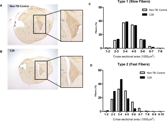
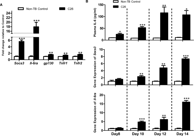
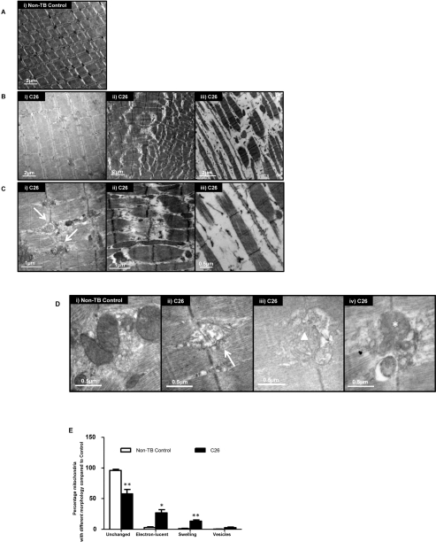
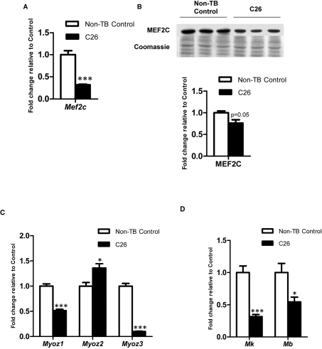
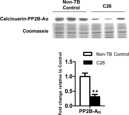
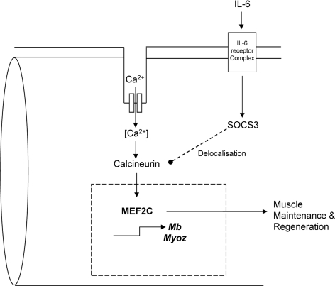
Cancer cachexia is a highly debilitating paraneoplastic disease observed in more than 50% of patients with advanced cancers and directly contributes to 20% of cancer deaths. Loss of skeletal muscle is a defining characteristic of patients with cancer cachexia and is associated with poor survival. The present study reveals the involvement of a myogenic transcription factor Myocyte Enhancer Factor (MEF) 2C in cancer-induced skeletal muscle wasting. Increased skeletal muscle mRNA expression of Suppressor of Cytokine Signaling (Socs) 3 and the IL-6 receptor indicative of active IL-6 signaling was seen in skeletal muscle of mice bearing the Colon 26 (C26) carcinoma. Loss of skeletal muscle structural integrity and distorted mitochondria were also observed using electron microscopy. Gene and protein expression of MEF2C was significantly downregulated in skeletal muscle from C26-bearing mice. MEF2C gene targets myozenin and myoglobin as well as myokinase were also altered during cachexia, suggesting dysregulated oxygen transport capacity and ATP regeneration in addition to distorted structural integrity. In addition, reduced expression of calcineurin was observed which suggested a potential pathway of MEF2C dysregulation. Together, these effects may limit sarcomeric contractile ability and also predispose skeletal muscle to structural instability; associated with muscle wasting and fatigue in cachexia.


Conflict of interest statement

The authors of this manuscript have no conflict of interest to declare.

Figures

Figure 1
Figure 1. Features of the C26 model of cachexia
Frozen sections of lower hindlimbs of (A) non-tumor-bearing (non-TB) control and (B) C26-bearing mice were immunohistochemically stained for MHC type I protein. Positive myofibers stained a relative dark brown compared to myofibers negative for the protein of interest. (C-D) The whole soleus muscle was subjected to myofiber cross-sectional area analysis to compare the proportion of myofiber type 1 (positively stained) and type 2 (negatively stained) in tumor-bearing mice versus non-TB controls. A more prominent reduction of bigger myofibers and a corresponding increase of smaller myofibers was seen in type 2 myofibers (n = 3).
Figure 2
Figure 2. mRNA expression of signaling molecules of the IL-6 signal transduction cascade
(A) Gene expression of Socs3, Il6ra, gp130, Tnfr1 and Tnfr2 were assessed as an indicator of inflammatory signaling (n = 4). Expression of all genes was increased at the endpoint in C26-bearing mice. (B) Longitudinal experiments demonstrated the rise of plasma IL-6 preceded the significant increase of Socs3 and Il-6ra at the transcript level. Data are presented as arithmetic means ± SEM. *p < 0.05, **p < 0.01, ***p < 0.001 compared to non-TB controls (n = 4 expect the non-TB groups on day 8 (n = 3) and day 14 (n = 2)).
Figure 3
Figure 3
Ultrastructural changes in GAS (A) Representative electron micrograph of muscle from non-tumor-bearing (non-TB) control mice. Magnification: × 8000. (B) Representative electron micrographs of muscle from C26-bearing mice. Magnification: × 8000 (i & ii), × 10000 (iii). (C) A higher magnification of muscle from C26-bearing mice. Vesicle-like structures (arrow); apparent tearing of myofiber (asterisk). Magnification: × 25000 (i & ii), × 30000 (iii). (D) Representative electron micrographs of muscle highlighting the morphologies of mitochondria. Electron-lucent areas (arrow); swelling (triangle); vesicle-like structures (asterisk). Magnification: × 40000 (i, ii & iv), × 30000 (iii). (E) Percentage of mitochondria with different morphologies in C26-bearing and non-TB mice. Data are presented as arithmetic means ± SEM. *p < 0.05, **p < 0.01 (n = 3). A reduced proportion of normal mitochondria and increased percentage of abnormal mitochondria with various changes were seen in C26-bearing mice compared to non-TB animals.
Figure 4
Figure 4. Expression of MEF2C and gene targets governing muscle structural integrity and energy homeostasis
Expression of MEF2C at (A) gene (n = 4) and (B) protein levels (n = 3). Expression of MEF2C target genes which govern (C) muscle structural intergrity and (D) energy homeostasis. Data are presented as arithmetic means ± SEM. *p < 0.05, ***p < 0.001 compared to non-tumor-bearing (non-TB) controls (n = 4). Expression of MEF2C was downregulated at both mRNA and protein levels. Altered mRNA expression was also seen in myozenins (myoz), myokinase (Mk) and myoglobin (Mb) which indicated disrupted muscle structure integrity and energy homeostasis in skeletal muscle during cancer cachexia.
Figure 5
Figure 5. Protein expression of calcineurin
The catalytic subunit of calcineurin was reduced at the protein level at endpoint cachexia. Data are presented as arithmetic means ± SEM. **p < 0.01 compared to non-tumor-bearing (non-TB) controls (n = 3).
Figure 6
Figure 6. A proposed model of IL-6 dependent loss of sarcomeric integrity a nd function involving MEF2C
Expression and activity of MEF2C are regulated by calcineurin. Chronic activation of IL-6 signaling in skeletal muscle results in an upregulated expression of SOCS3. SOCS3 has been shown to delocalize calcineurin from its usual Z-line position to the periphery of a myofiber. This might affect its function and hence impact on the downstream targets like MEF2C. Since MEF2C is a key regulator of many myogenic and energy homeostatic molecules, any perturbation in its activity could greatly affect muscle performance and integrity. Such alterations may accelerate muscle breakdown and disintegration of the tissue contributing to fatigue and weakness during cancer cachexia.

Similar articles

Cited by

References

    1. Tisdale MJ. Molecular pathways leading to cancer cachexia. Physiology (Bethesda) 2005;20:340–348. - PubMed
    1. Evans WJ, Morley JE, Argiles J, Bales C, Baracos V, Guttridge D, Jatoi A, Kalantar-Zadeh K, Lochs H, Mantovani G, Marks D, Mitch WE, Muscaritoli M, Najand A, Ponikowski P, Rossi Fanelli F, et al. Cachexia: a new definition. Clin Nutr. 2008;27:793–799. - PubMed
    1. Zhou X, Wang JL, Lu J, Song Y, Kwak KS, Jiao Q, Rosenfeld R, Chen Q, Boone T, Simonet WS, Lacey DL, Goldberg AL, Han HQ. Reversal of cancer cachexia and muscle wasting by ActRIIB antagonism leads to prolonged survival. Cell. 2010;142:531–543. - PubMed
    1. Guttridge DC, Mayo MW, Madrid LV, Wang CY, Baldwin AS., Jr. NF-kappaB-induced loss of MyoD messenger RNA: possible role in muscle decay and cachexia. Science. 2000;289:2363–2366. - PubMed
    1. Carson JA, Baltgalvis KA. Interleukin 6 as a key regulator of muscle mass during cachexia. Exerc Sport Sci Rev. 2010;38:168–176. - PMC - PubMed

Publication types

MeSH terms