. 2012 May 15;302(10):G1111-32.
doi: 10.1152/ajpgi.00519.2011. Epub 2012 Feb 23.

Activation of two distinct Sox9-EGFP-expressing intestinal stem cell populations during crypt regeneration after irradiation

Affiliations

Activation of two distinct Sox9-EGFP-expressing intestinal stem cell populations during crypt regeneration after irradiation

Laurianne Van Landeghem et al. Am J Physiol Gastrointest Liver Physiol. .

Abstract

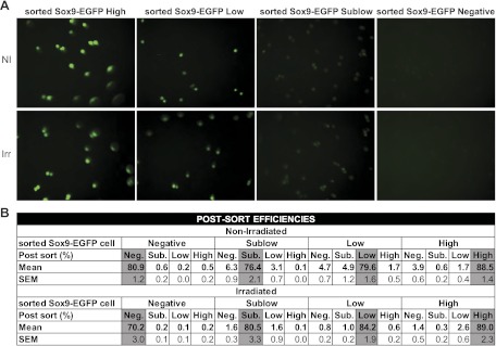
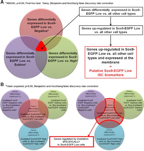
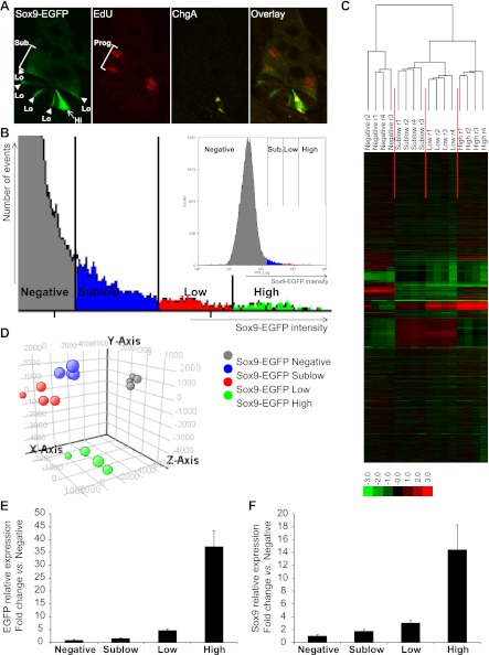
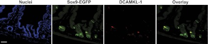
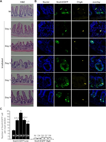
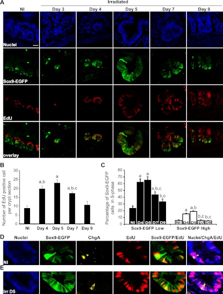
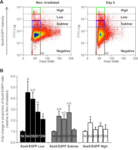
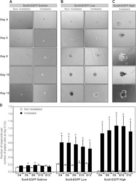
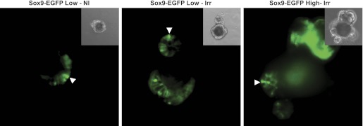
Recent identification of intestinal epithelial stem cell (ISC) markers and development of ISC reporter mice permit visualization and isolation of regenerating ISCs after radiation to define their functional and molecular phenotypes. Previous studies in uninjured intestine of Sox9-EGFP reporter mice demonstrate that ISCs express low levels of Sox9-EGFP (Sox9-EGFP Low), whereas enteroendocrine cells (EEC) express high levels of Sox9-EGFP (Sox9-EGFP High). We hypothesized that Sox9-EGFP Low ISCs would expand after radiation, exhibit enhanced proliferative capacities, and adopt a distinct gene expression profile associated with rapid proliferation. Sox9-EGFP mice were given 14 Gy abdominal radiation and studied between days 3 and 9 postradiation. Radiation-induced changes in number, growth, and transcriptome of the different Sox9-EGFP cell populations were determined by histology, flow cytometry, in vitro culture assays, and microarray. Microarray confirmed that nonirradiated Sox9-EGFP Low cells are enriched for Lgr5 mRNA and mRNAs enriched in Lgr5-ISCs and identified additional putative ISC markers. Sox9-EGFP High cells were enriched for EEC markers, as well as Bmi1 and Hopx, which are putative markers of quiescent ISCs. Irradiation caused complete crypt loss, followed by expansion and hyperproliferation of Sox9-EGFP Low cells. From nonirradiated intestine, only Sox9-EGFP Low cells exhibited ISC characteristics of forming organoids in culture, whereas during regeneration both Sox9-EGFP Low and High cells formed organoids. Microarray demonstrated that regenerating Sox9-EGFP High cells exhibited transcriptomic changes linked to p53-signaling and ISC-like functions including DNA repair and reduced oxidative metabolism. These findings support a model in which Sox9-EGFP Low cells represent active ISCs, Sox9-EGFP High cells contain radiation-activatable cells with ISC characteristics, and both participate in crypt regeneration.


Figures

Fig. 1.
Fig. 1.
Postsort analysis of the different fluorescence-activated cell sorting (FACS) isolated Sox9-EGFP cell populations. A: representative photographs of the 4 Sox9-EGFP cell fractions isolated by FACS demonstrate that they express appropriate intensities of enhanced green fluorescence protein (EGFP) fluorescence (magnification ×40). B: data from flow cytometry used to assess the postsort efficiencies and potential cross-contamination of cells that do not express Sox9-EGFP and those expressing sublow, low, and high levels of Sox9-EGFP (Sox9-EGFP Negative, Sublow, Low, and High cells, respectively) with other Sox9-EGFP cell populations. Data are expressed as means and SE of the postsort percentages of corresponding cells collected for each sort (n ≥ 9 in each group). Note that each sorted cell population contains more than 70% of the appropriate cell type and presents minimal contamination with other cells. Importantly, the postsort efficiencies and cross-contamination values do not significantly differ in cells from nonirradiated and irradiated mice.
Fig. 2.
Fig. 2.
Schematization of approaches used to analyze the microarray data. A: analysis approach used to identify new putative Sox9-EGFP Low intestinal epithelial stem cell (ISC) biomarkers. Briefly, statistical analysis identified genes significantly differentially regulated in Sox9-EGFP Low cells vs. Negative cells (red), Sublow cells (purple), or High cells (green) (ANOVA; P < 0.05; Tukey; Benjamini-Hochberg multiple testing comparison). By selecting genes that were common to each of these 3 analyses, we isolated genes that were differentially regulated in Sox9-EGFP Low cells vs. all other cell types. Genes that were upregulated in Sox9-EGFP Low ISCs vs. all other cell types and that encode membrane proteins were selected as putative Sox9-EGFP Low ISC biomarkers (see Tables 5 and 6). This same analysis strategy was used to identify new putative Sox9-EGFP Sublow progenitor biomarkers (see Tables 5 and 6). B: analysis approach used to identify genes whose expression was regulated by irradiation specifically in Sox9-EGFP Low ISCs. Briefly, statistical analysis identified genes significantly differentially regulated in Sox9-EGFP Low cells isolated from mice at day 5 postirradiation vs. Sox9-EGFP Low cells from nonirradiated mice (red). Similar analysis was performed for Sox9-EGFP Negative (purple), Sublow cells (green), and High cells (blue) (t-test unpaired; P < 0.05; Benjamini-Hochberg multiple testing comparison). We then selected genes that were regulated after irradiation exclusively in Sox9-EGFP Low ISCs. This same analysis strategy was used to identify genes regulated by irradiation specifically in Sox9-EGFP Sublow or High cells.
Fig. 3.
Fig. 3.
Sox9-EGFP transgene marks 4 different cell populations that exhibit distinct gene expression signatures. A: confocal microscopy on a jejunal crypt from a nonirradiated Sox9-EGFP mouse illustrates that the Sox9-EGFP transgene (green) is expressed at different levels (Hi, high/intense staining, arrow; Lo, low/moderate staining, arrowhead; Sub., sublow/very low staining). Note that in this and subsequent histological figures, we use the “High, Low, and Sublow” terminology to describe cells with intense, moderate, and very low intensities of Sox9-EGFP fluorescence, respectively. 5-Ethynyl-2′deoxyuridine (EdU)-positive cells (red) are predominantly located in the progenitor (Prog.) compartment and express sublow levels of Sox9-EGFP. Chromogranin-A (ChgA; yellow) is localized to Sox9-EGFP High cells (magnification ×40). B: representative graph illustrates gating used for flow cytometry and FACS-based isolation of Sox9-EGFP-expressing cells from single cell preparations of IECs from nonirradiated mouse jejunum. Four levels of expression of the transgene (Sox9-EGFP intensity) can be distinguished: Negative (gray), Sublow (blue), Low (red), and High (green). Hierarchical clustering (C) and principal component analysis (PCA; D) on gene expression data from the 4 Sox9-EGFP cell populations isolated from 4 independent nonirradiated Sox9-EGFP mice reveal 4 distinct and consistent gene expression signatures (hierarchical clustering on genes and conditions, Spearman rank correlation, single linkage, r, replicate; PCA on conditions, 4 principal components, mean centered). E: real-time quantitative PCR (qPCR) data showed that the expression profile of EGFP was consistent with the 4 different Sox9-EGFP sorted groups (i.e., Negative, Sublow, Low, and High). Data are expressed as fold change (mean ± SE) vs. Sox9-EGFP Negative cells (n = 6). F: real-time qPCR data demonstrated that the expression of the endogenous Sox9 gene matched the expression levels of the Sox9-EGFP transgene in the different sorted cell populations. Data are expressed as fold change (mean ± SE) vs. Sox9-EGFP Negative cells (n = 6).
Fig. 4.
Fig. 4.
DCAMKL-1 colocalizes with Sox9-EGFP High cells. Immunostaining for Sox9-EGFP (green) and DCAMKL-1 (red) demonstrates that in intestinal epithelial crypts, DCAMKL-1 colocalizes with high levels of Sox9-EGFP [nuclei staining, 4,6-diamidino-2-phenylindole (DAPI), blue; scale bar 50 μm].
Fig. 5.
Fig. 5.
Sox9-EGFP Low ISCs expand during crypt regeneration following radiation. A: hematoxylin and eosin (H&E) staining of jejunum from nonirradiated (NI) and irradiated mice at days 3, 4, 5, 7, and 9 after 14 Gy abdominal irradiation demonstrates crypt loss at day 3, formation of regenerating microcolonies at day 4; crypt hyperplasia at days 5 (D5) and 7 (D7) and a trend toward baseline crypt and villus morphology by day 9. Scale bar 100 μm. B: immunostaining for Sox9-EGFP (green) and ChgA (yellow) demonstrates that regenerating microcolonies and hyperplastic crypts are highly enriched in Sox9-EGFP Low cells whereas Sox9-EGFP High cells colabeled with ChgA did not expand after radiation (nuclei staining, DAPI, blue; confocal images; scale bar 25 μm). C: histogram shows means ± SE for the number of Sox9-EGFP High or Low cells per crypt section. At least 30 crypt sections were analyzed per animal by 3 independent blinded observers (n ≥ 3 independent mice at each time point; aP < 0.001 vs. nonirradiated; bP < 0.001 vs. day 5; cP < 0.05 vs. day 9).
Fig. 6.
Fig. 6.
Increase in proliferating Sox9-EGFP Low and High cells during crypt regeneration. A: EdU (red) and Sox9-EGFP (green) colocalization reveals that a large proportion of cells in regenerating microcolonies at day 4 and hyperplastic crypts observed at days 5 and 7 were positive for EdU and illustrates increased numbers of Sox9-EGFP Low cells that are EdU positive during crypt regeneration (nuclei staining, DAPI, blue; scale bar 25 μm). B: histogram shows mean ± SE of the number of EdU-positive cells per crypt section. At least 30 crypts were studied per animal per time point (n = 3 at each time point; aP < 0.01 vs. nonirradiated; bP < 0.05 vs. day 9; cP < 0.05 vs. day 5). C: means ± SE of the percentage of Sox9-EGFP Low (black bars) and High (white bars) cells colabeled with EdU. Early phases of regeneration after radiation involve significant increases in the proportion of Sox9-EGFP Low and High cells in S-phase (n = 3 at each time point; aP < 0.05 vs. nonirradiated; bP < 0.05 vs. day 4; cP < 0.05 vs. day 5). D and E: confocal images of a crypt from jejunum of a nonirradiated Sox9-EGFP mouse (D) and at 5 days postirradiation (Irr D5) (E) illustrating Sox9-EGFP High cells (green) expressing ChgA (yellow) and colabeled with EdU (red) (nuclei staining, DAPI, blue; magnification ×40).
Fig. 7.
Fig. 7.
Irradiation induces distinct changes in the proportion of Sox9-EGFP High, Low, and Sublow cells. A: representative flow cytometry data show the proportion of cells expressing High (green rectangle), Low (red rectangle), and Sublow (blue rectangle) levels of Sox9-EGFP (Sox9-EGFP intensity) isolated from a nonirradiated mouse or at day 4 postradiation. Note the marked and obvious increase in the proportion of Sox9-EGFP Low and Sublow cells at day 4 after radiation. B: histograms show the quantification of the flow cytometry data expressed as mean fold change ± SE in the proportion of Sox9-EGFP High, Low or Sublow cells at each time point postradiation vs. nonirradiated controls (n ≥ 3 at each time point; aP < 0.05 vs. nonirradiated; bP < 0.05 vs. day 9).
Fig. 8.
Fig. 8.
Sox9-EGFP Low and High cells isolated at 5 days postirradiation possess enhanced ability to form organoids in vitro. Organoid cultures were performed on FACS-isolated cells from nonirradiated mice or at day 5 postradiation and were monitored at 2-day intervals between day 4 (D4) and day 12 (D12) after plating. Representative photographs show Sox9-EGFP Sublow cells from nonirradiated or irradiated mice (A), Sox9-EGFP Low cells from nonirradiated or irradiated mice (B), and Sox9-EGFP High cells isolated at day 5 postradiation (C) (bright field; scale bar 200 μm). D: histograms show quantitative data (mean ± SE) for the number of organoids formed by the 3 different Sox9-EGFP-sorted cell populations at each time point divided by the initial number of plated cells ×100 (n ≥ 3; aP < 0.05 vs. nonirradiated). Sox9-EGFP Low and High cells isolated from irradiated mice exhibited significantly greater ability to form organoids compared with nonirradiated controls.
Fig. 9.
Fig. 9.
Organoids grown from nonirradiated and irradiated Sox9-EGFP Low cells and from Sox9-EGFP High cells isolated at 5 days postirradiation each express both low and high levels of Sox9-EGFP. Representative photographs shows EGFP fluorescence of organoids obtained from nonirradiated Sox9-EGFP Low cells (left; Sox9-EGFP Low-NI), Sox9-EGFP Low cells isolated at 5 days postradiation (middle; Sox9-EGFP Low-Irr), and Sox9-EGFP High cells isolated at 5 days postradiation (right, Sox9-EGFP High-Irr). Organoids contained discrete cells expressing Low/moderate or High/intense levels of Sox9-EGFP. Arrowheads show cells expressing High/intense levels of the Sox9-EGFP transgene. Note: High-intensity green in upper right corner of the photograph illustrating EGFP fluorescence of organoid obtained from Sox9-EGFP High cells isolated at 5 days postradiation is autofluorescence from an aggregate of dead cells.

References

    1. Alarcon VB. Cell polarity regulator PARD6B is essential for trophectoderm formation in the preimplantation mouse embryo. Biol Reprod 83: 347–358, 2010 - PMC - PubMed
    1. Barker N, van de Wetering M, Clevers H. The intestinal stem cell. Genes Dev 22: 1856–1864, 2008 - PMC - PubMed
    1. Barker N, van Es JH, Kuipers J, Kujala P, van den Born M, Cozijnsen M, Haegebarth A, Korving J, Begthel H, Peters PJ, Clevers H. Identification of stem cells in small intestine and colon by marker gene Lgr5. Nature 449: 1003–1007, 2007 - PubMed
    1. Batlle E, Henderson JT, Beghtel H, van den Born MM, Sancho E, Huls G, Meeldijk J, Robertson J, van de Wetering M, Pawson T, Clevers H. Beta-catenin and TCF mediate cell positioning in the intestinal epithelium by controlling the expression of EphB/ephrinB. Cell 111: 251–263, 2002 - PubMed
    1. Bhanja P, Saha S, Kabarriti R, Liu L, Roy-Chowdhury N, Roy-Chowdhury J, Sellers RS, Alfieri AA, Guha C. Protective role of R-spondin1, an intestinal stem cell growth factor, against radiation-induced gastrointestinal syndrome in mice. PLoS One 4: e8014, 2009 - PMC - PubMed

Publication types

MeSH terms