. 2012 Jun 1;302(11):G1347-55.
doi: 10.1152/ajpgi.00013.2012. Epub 2012 Feb 23.

Esophageal functional impairments in experimental eosinophilic esophagitis

Affiliations

Esophageal functional impairments in experimental eosinophilic esophagitis

Parm Mavi et al. Am J Physiol Gastrointest Liver Physiol. .

Abstract

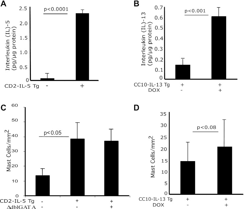
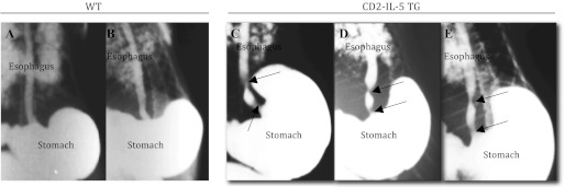
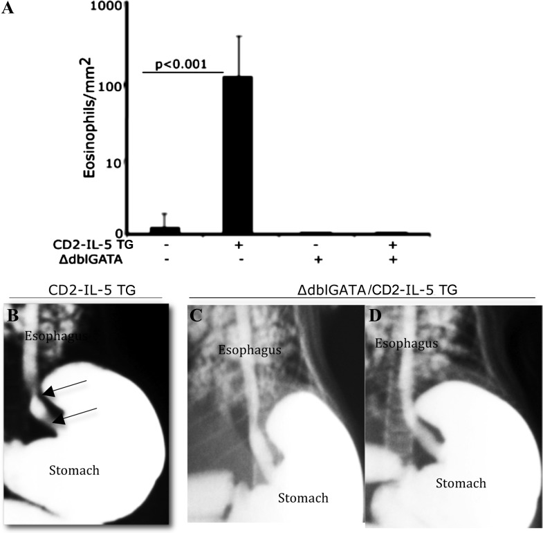
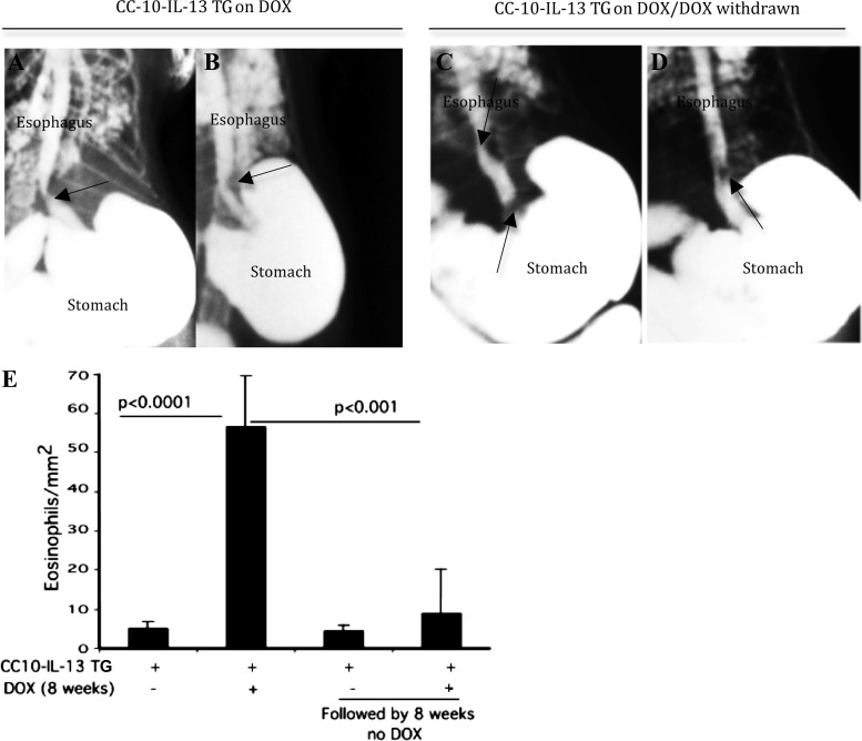
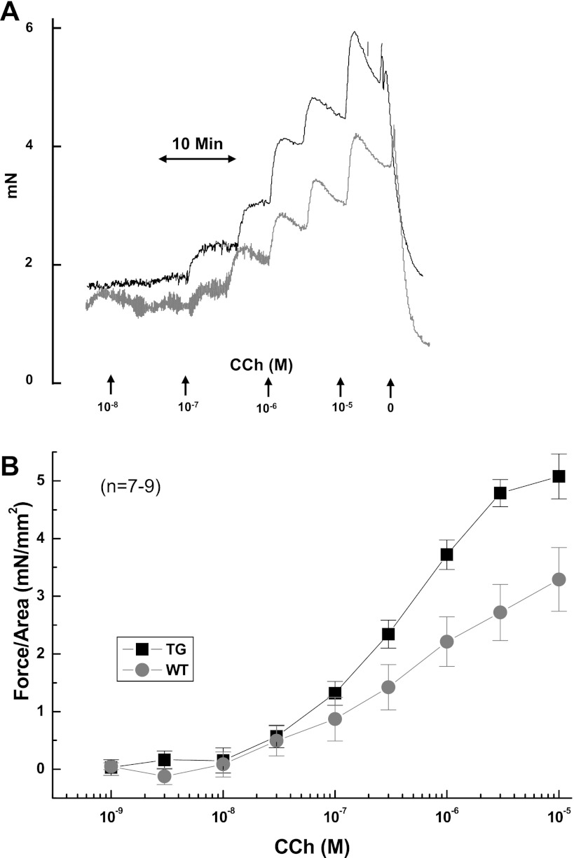
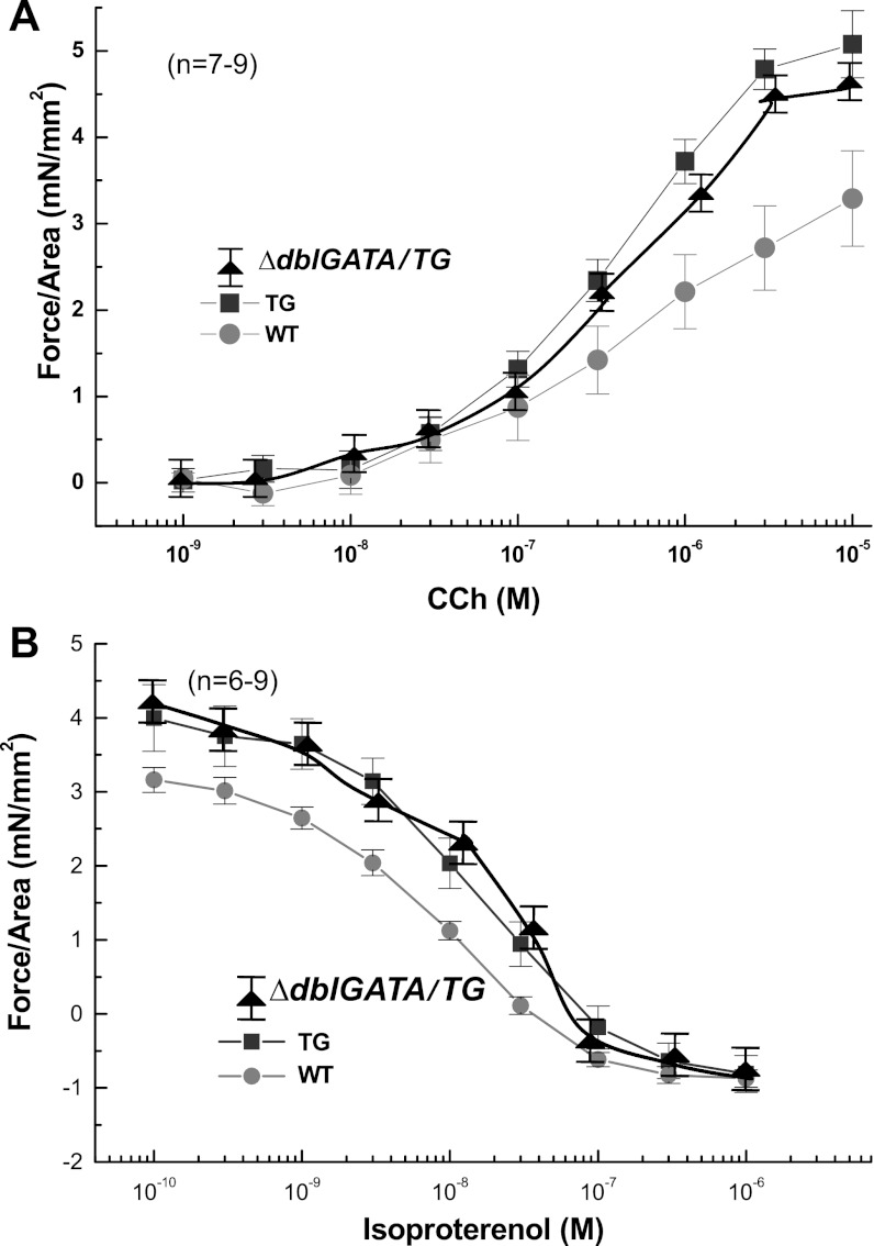
Eosinophilic esophagitis (EoE) is an emerging chronic esophageal disease. Despite the increasing diagnosis of EoE globally, the causes of EoE and other esophageal eosinophilic disorders are not clearly understood. EoE pathology includes accumulation of inflammatory cells (e.g., eosinophils, mast cells), characteristic endoscopic features (e.g., furrows, the formation of fine concentric mucosal rings, exudates), and functional impairments (e.g., esophageal stricture, dysmotility). We hypothesized that the esophageal structural pathology and functional impairments of EoE develop as a consequence of the effector functions of the accumulated inflammatory cells. We analyzed eosinophils (anti-major basic protein immunostaining), esophageal stricture (X-ray barium swallowing), and esophageal motility (isometric force) in two established transgenic murine models of EoE (CD2-IL-5 and rtTA-CC10-IL-13) and a novel eosinophil-deficient model (ΔdblGATA/CD2-IL-5). Herein, we show the following: 1) CD2-IL-5 and doxycycline (DOX)-induced rtTA-CC10-IL-13 mice have chronic eosinophilic and mast cell esophageal inflammation; 2) eosinophilic esophageal inflammation promotes esophageal stricture in both transgenic murine models; 3) the eosinophil-deficient ΔdblGATA/CD-2-IL-5 mice were protected from the induction of stricture, whereas the eosinophil-competent CD2-IL-5 mice develop esophageal stricture; 4) esophageal stricture is not reversible in DOX-induced rtTA-CC10-IL-13 mice (8 wk DOX followed by 8 wk no-DOX); and 5) IL-5 transgene-induced (CD2-IL-5) EoE evidences esophageal dysmotility (relaxation and contraction) that is independent of the eosinophilic esophageal inflammation: CD2-IL-5 and ΔdblGATA/CD2-IL-5 mice have comparable esophageal dysmotility. Collectively, our present study directly implicates chronic eosinophilic inflammation in the development of the esophageal structural impairments of experimental EoE.


Figures

Fig. 1.
Fig. 1.
IL-5, IL-13, and mast cell induction in the esophagus. Protein levels of IL-5 and IL-13 from esophageal tissue of wild-type (WT), CD2-IL-5, and uninduced and doxycycline (DOX)-induced rtTA-CC10-IL-13 mice were measured by performing ELISA. Levels of IL-5 in 12-wk-old WT and in CD2-IL-5 mice and of IL-13 in 8-wk-old WT and in rtTA-CC-10-IL-13 mice are shown (A and B). The level of mast cells in the esophagus of WT, CD2-IL-5, ΔdblGATA/CD2-IL-5, and DOX- and no-DOX-treated rtTA-CC10-IL-13 mice were quantified by performing morphometric analysis following chloroacetate tissue staining (C and D). The results are the summary of 3 independent experiments, reported as means ± SE with n = 6 mice for each group. The statistical P values are provided in each figure.
Fig. 2.
Fig. 2.
Esophageal stricture in IL-5-induced esophageal inflammation. The WT and CD2-IL-5 mice were given 10% barium sulfate solution orally via a 5-ml syringe. Mice were allowed to slowly swallow ∼3–4 ml of the solution and then anesthetized with isoflurane. X-ray analysis was performed on the whole body. Radiological barium-swallowing experiments indicated esophageal stricture in CD2-IL-5 mice develops in response to IL-5-induced chronic inflammation in the esophagus. Representative radiophotographs of WT (A and B) and CD2-IL-5 (C–E) mice are shown. Esophageal stricture is marked by the arrows for the CD2-IL-5 mice; no stricture is visible in WT mice. For WT and CD2-IL5 mice, n = 8–10/group. TG, transgenic.
Fig. 3.
Fig. 3.
GATA-1 deficiency in CD2 promoter-driven IL-5 transgenic (TG) mice prevents esophageal eosinophilia and stricture. Esophageal eosinophils in wild-type (WT) and CD2-IL-5 (IL-5 TG), ΔdblGATA, and ΔdblGATA/CD2-IL-5 TG mice were examined. The morphometric analysis indicated an increase in the number of esophageal eosinophils in CD2-IL-5 mice compared with WT, ΔdblGATA (no IL-5 transgene), and ΔdblGATA/CD2-IL-5 mice (A). Both ΔdblGATA and ΔdblGATA/CD2-IL-5 showed no esophageal eosinophils. Furthermore, barium-swallowing experiments showed that ΔdblGATA/CD2-IL-5 TG mice are protected from the development of esophageal stricture. Representative X-ray radiophotographs of CD-IL-5 (B, esophageal stricture is marked by the arrows) and ΔdblGATA/CD2-IL-5 (C and D) mice are shown. Data are expressed as means ± SE, n = 10 mice/group.
Fig. 4.
Fig. 4.
Esophageal stricture is irreversible even after reducing the eosinophilic inflammation in mice. Esophageal eosinophilic inflammation was induced in DOX-regulated IL-13 TG mice (rtTA-CC10-IL-13) following an 8-wk regimen of DOX food. One group of mice was examined for the development of esophageal stricture after the 8-wk DOX food regimen (representative radiophotographs; A and B). The second group of mice was switched to normal (no-DOX) food for 8 wk and then examined for stricture (representative radiophotographs; C and D). Both groups of mice showed esophageal stricture (arrows). A high degree of esophageal eosinophilia is shown in the 8-wk DOX-exposed group of rtTA-CC10-IL-13 mice compared with the significantly reduced esophageal eosinophilia in the DOX withdrawn (after 8 wk) group of rtTA-CC10-IL-13 mice (E). The 16-wk rTA-CC10-IL-13 mice with no-DOX food show a baseline eosinophilia that is not statistically different from that of the 8 wk on DOX followed by 8 wk no-DOX food given rTA-CC10-IL-13 mice (E). Data are shown as means ± SD, n = 8 mice/group.
Fig. 5.
Fig. 5.
Carbachol (CCh)-induced esophageal muscle contraction impairment in a murine model of eosinophilic esophagitis. A representative kinetic tracing of the dose-dependent contraction of esophageal muscles from wild-type (WT) (gray line) and CD2-IL-5 (TG; black line) mice in response to increasing concentrations of carbachol is shown (A). The concentration-dependent, carbachol-induced esophageal muscle contraction of esophageal longitudinal muscles from WT (gray circles with line) and CD2-IL-5 (TG, black squares with line) mice was measured by adding increasing concentrations of carbachol, as indicated in the figure (B). Data are given as means ± SE, n = 6 mice/group, P < 0.001.
Fig. 6.
Fig. 6.
Isoproterenol (ISO)-induced esophageal muscle relaxation impairment in a murine model of eosinophilic esophagitis. A representative kinetic tracing of the dose-dependent relaxation of carbachol-precontracted (105 M) esophageal smooth muscles from WT (gray line) and CD2-IL-5 (TG; black line) mice in response to increasing concentrations of isoproterenol is shown (A). The isoproterenol-induced relaxation of carbachol-precontracted esophageal muscles from WT (gray circles with line) and CD2-IL-5 (TG, black squares with line) mice was measured by adding increasing concentrations of isoproterenol, as indicated in the figure (B). Data are given as means ± SE, n = 6 mice/group, P < 0.001.
Fig. 7.
Fig. 7.
Esophageal motility impairment is independent of eosinophilic inflammation. The concentration-dependent, carbachol-induced contraction of esophageal longitudinal muscle and isoproterenol-induced relaxation of carbachol-precontracted (105 M) esophageal longitudinal muscle contraction was measured in WT, CD2-IL-5, and ΔdblGATA/CD2-IL-5 mice by adding increasing concentrations of carbachol or isoproterenol, as indicated (A and B). Esophageal muscle contraction and relaxation in CD2-IL-5 mice is impaired compared with WT mice; comparable esophageal motility impairment is shown between CD2-IL-5 TG and ΔdblGATA/CD2-IL-5 TG mice (A and B). Data are given as means ± SE, n = 6 mice/group, P < 0.001.

References

    1. Attwood SE, Smyrk TC, Demeester TR, Jones JB. Esophageal eosinophilia with dysphagia. A distinct clinicopathologic syndrome. Dig Dis Sci 38: 109–116, 1993 - PubMed
    1. Blanchard C, Wang N, Stringer KF, Mishra A, Fulkerson PC, Abonia JP, Jameson SC, Kirby C, Konikoff MR, Collins MH, Cohen MB, Akers R, Hogan SP, Assa'ad AH, Putnam PE, Aronow BJ, Rothenberg ME. Eotaxin-3 and a uniquely conserved gene-expression profile in eosinophilic esophagitis. J Clin Invest 116: 536–547, 2006 - PMC - PubMed
    1. Fox VL, Nurko S, Furuta GT. Eosinophilic esophagitis: It's not just kid's stuff. Gastrointest Endosc 56: 260–270, 2002 - PubMed
    1. Furuta GT, Liacouras CA, Collins MH, Gupta SK, Justinich C, Putnam PE, Bonis P, Hassall E, Straumann A, Rothenberg ME. Eosinophilic esophagitis in children and adults: a systematic review and consensus recommendations for diagnosis and treatment. Gastroenterology 133: 1342–1363, 2007 - PubMed
    1. Gleich GJ, Adolphson CR. The eosinophilic leukocyte: structure and function. Adv Immunol 39: 177–253, 1986 - PubMed

Publication types

MeSH terms