

A bimodal influence of thyroid hormone on cerebellum oligodendrocyte differentiation
- PMID: 22361821
- PMCID: PMC5417134
- DOI: 10.1210/me.2011-1316
A bimodal influence of thyroid hormone on cerebellum oligodendrocyte differentiation
Abstract
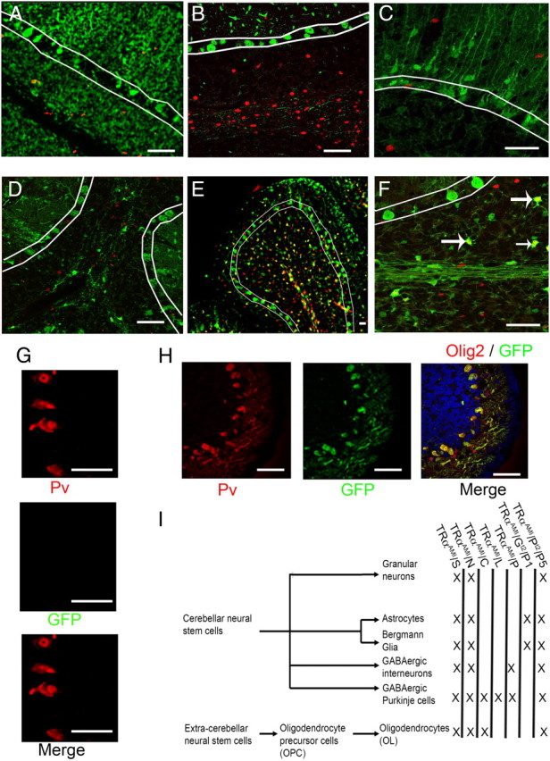
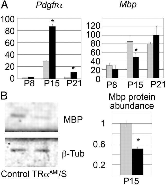
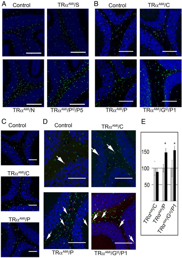
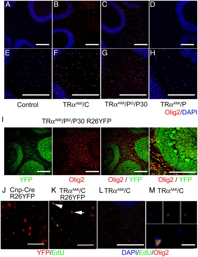
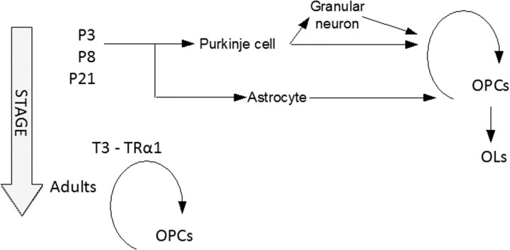
Thyroid hormone (T(3)) can trigger a massive differentiation of cultured oligodendrocytes precursor cells (OPC) by binding the nuclear T(3) receptor α1 (TRα1). Whether this reflects a physiological function of TRα1 remains uncertain. Using a recently generated mouse model, in which CRE/loxP recombination is used to block its function, we show that TRα1 acts at two levels for the in vivo differentiation of OPC in mouse cerebellum. At the early postnatal stage, it promotes the secretion of several neurotrophic factors by acting in Purkinje neurons and astrocytes, defining an environment suitable for OPC differentiation. At later stages, TRα1 acts in a cell-autonomous manner to ensure the complete arrest of OPC proliferation. These data explain contradictory observations made on various models and outline the importance of T(3) signaling both for synchronizing postnatal neurodevelopment and restraining OPC proliferation in adult brain.
Figures
Similar articles
-
Purkinje cells and Bergmann glia are primary targets of the TRα1 thyroid hormone receptor during mouse cerebellum postnatal development.Development. 2014 Jan;141(1):166-75. doi: 10.1242/dev.103226. Development. 2014. PMID: 24346699
-
The thyroid hormone receptor alpha1 protein is expressed in embryonic postmitotic neurons and persists in most adult neurons.Mol Endocrinol. 2010 Oct;24(10):1904-16. doi: 10.1210/me.2010-0175. Epub 2010 Aug 25. Mol Endocrinol. 2010. PMID: 20739404 Free PMC article.
-
Thyroid hormone induces cerebellar Purkinje cell dendritic development via the thyroid hormone receptor alpha1.J Neurosci. 2003 Nov 19;23(33):10604-12. doi: 10.1523/JNEUROSCI.23-33-10604.2003. J Neurosci. 2003. PMID: 14627645 Free PMC article.
-
[Thyroid hormone and cerebellum development: direct and indirect effects?].Ann Endocrinol (Paris). 2011 Apr;72(2):99-102. doi: 10.1016/j.ando.2011.03.012. Epub 2011 Apr 20. Ann Endocrinol (Paris). 2011. PMID: 21511240 Review. French.
-
Oligodendrocyte development and thyroid hormone.J Neurobiol. 1999 Sep 15;40(4):497-512. doi: 10.1002/(sici)1097-4695(19990915)40:4<497::aid-neu7>3.0.co;2-#. J Neurobiol. 1999. PMID: 10453052 Review.
Cited by
-
Involvement of Thyroid Hormones in Brain Development and Cancer.Cancers (Basel). 2021 May 30;13(11):2693. doi: 10.3390/cancers13112693. Cancers (Basel). 2021. PMID: 34070729 Free PMC article. Review.
-
Cerebellar abnormalities in mice lacking type 3 deiodinase and partial reversal of phenotype by deletion of thyroid hormone receptor α1.Endocrinology. 2013 Jan;154(1):550-61. doi: 10.1210/en.2012-1738. Epub 2012 Nov 16. Endocrinology. 2013. PMID: 23161871 Free PMC article.
-
Thyroid hormone role on cerebellar development and maintenance: a perspective based on transgenic mouse models.Front Endocrinol (Lausanne). 2014 May 20;5:75. doi: 10.3389/fendo.2014.00075. eCollection 2014. Front Endocrinol (Lausanne). 2014. PMID: 24904526 Free PMC article. Review.
-
Thyroid hormone triggers the developmental loss of axonal regenerative capacity via thyroid hormone receptor α1 and krüppel-like factor 9 in Purkinje cells.Proc Natl Acad Sci U S A. 2012 Aug 28;109(35):14206-11. doi: 10.1073/pnas.1119853109. Epub 2012 Aug 13. Proc Natl Acad Sci U S A. 2012. PMID: 22891348 Free PMC article.
-
Influence of hormones in multiple sclerosis: focus on the most important hormones.Metab Brain Dis. 2023 Mar;38(3):739-747. doi: 10.1007/s11011-022-01138-7. Epub 2023 Jan 3. Metab Brain Dis. 2023. PMID: 36595158 Review.
References
-
- Calver AR , Hall AC , Yu WP , Walsh FS , Heath JK , Betsholtz C , Richardson WD. 1998. Oligodendrocyte population dynamics and the role of PDGF in vivo. Neuron 20:869–882 - PubMed
-
- Barres BA , Raff MC , Gaese F , Bartke I , Dechant G , Barde YA. 1994. A crucial role for neurotrophin-3 in oligodendrocyte development. Nature 367:371–375 - PubMed
