. 2013 Jan;97(1):181-93.
doi: 10.1007/s00253-012-3943-1. Epub 2012 Feb 25.

4,6-α-Glucanotransferase activity occurs more widespread in Lactobacillus strains and constitutes a separate GH70 subfamily

Affiliations

4,6-α-Glucanotransferase activity occurs more widespread in Lactobacillus strains and constitutes a separate GH70 subfamily

Hans Leemhuis et al. Appl Microbiol Biotechnol. 2013 Jan.

Abstract

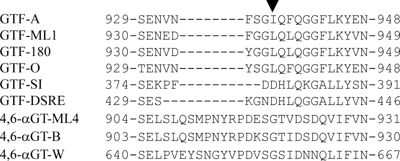
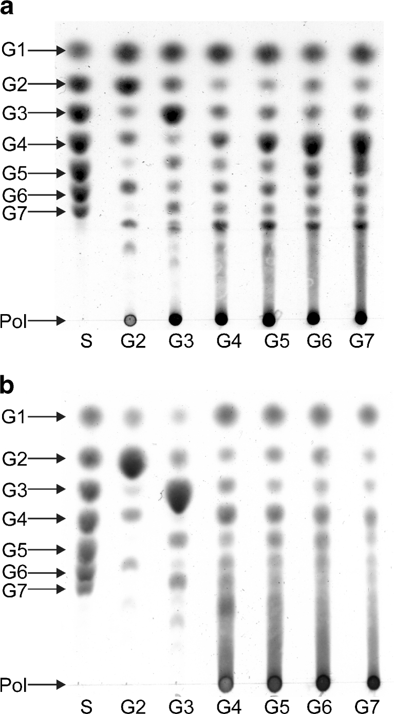
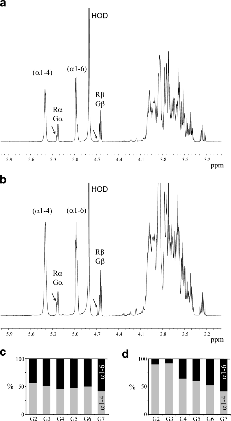
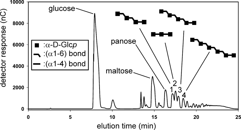
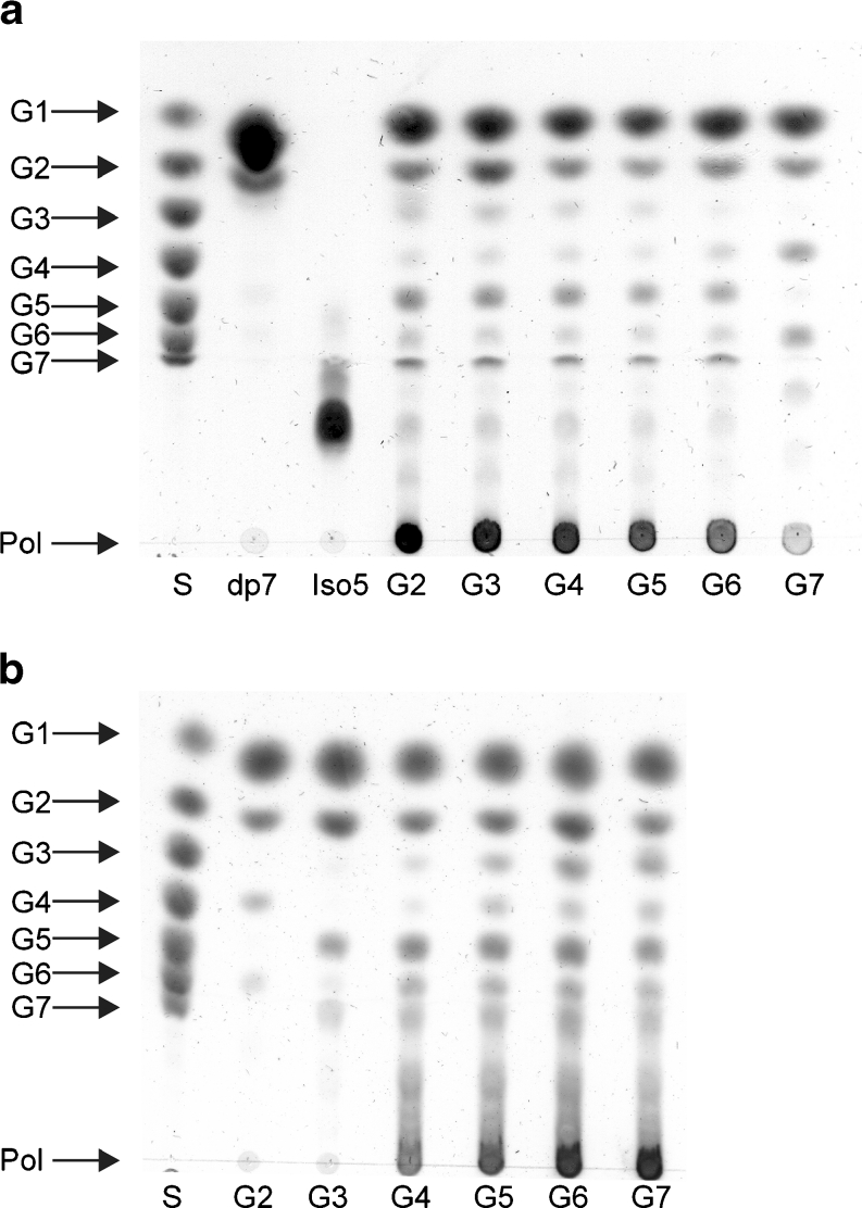
Family 70 glycoside hydrolase glucansucrase enzymes exclusively occur in lactic acid bacteria and synthesize a wide range of α-D-glucan (abbreviated as α-glucan) oligo- and polysaccharides. Of the 47 characterized GH70 enzymes, 46 use sucrose as glucose donor. A single GH70 enzyme was recently found to be inactive with sucrose and to utilize maltooligosaccharides [(1→4)-α-D-glucooligosaccharides] as glucose donor substrates for α-glucan synthesis, acting as a 4,6-α-glucanotransferase (4,6-αGT) enzyme. Here, we report the characterization of two further GH70 4,6-αGT enzymes, i.e., from Lactobacillus reuteri strains DSM 20016 and ML1, which use maltooligosaccharides as glucose donor. Both enzymes cleave α1→4 glycosidic linkages and add the released glucose moieties one by one to the non-reducing end of growing linear α-glucan chains via α1→6 glycosidic linkages (α1→4 to α1→6 transfer activity). In this way, they convert pure maltooligosaccharide substrates into linear α-glucan product mixtures with about 50% α1→6 glycosidic bonds (isomalto/maltooligosaccharides). These new α-glucan products may provide an exciting type of carbohydrate for the food industry. The results show that 4,6-αGTs occur more widespread in family GH70 and can be considered as a GH70 subfamily. Sequence analysis allowed identification of amino acid residues in acceptor substrate binding subsites +1 and +2, differing between GH70 GTF and 4,6-αGT enzymes.


Figures

Fig. 1
Fig. 1
The loop connecting the domains IV and B is longer in 4,6-αGTs then in GTFs. The sequence alignment shows the region around the subsite +1 residue L938 (indicated by the triangle) of GTF-180. The sequences used are as follows: GTF-A (Q5SBL9) of Lactobacillus reuteri 121; GTF-180 (Q5SBN3) of L. reuteri 180; GTF-O (Q4JLC7) of L. reuteri ATCC 55730; GTF-ML1 (Q5SBN0) of L. reuteri ML1; GTF-DSRE (Q8G9Q2) of Leuconostoc mesenteroides NRRL B-1299; GTF-SI (P13470) of Streptococcus mutans GS-5; 4,6-αGT-B (Q5SBM0) of L. reuteri 121; 4,6-αGT-W (A5VL73) of L. reuteri DSM20016; 4,6-αGT-ML4 (Q5SBN1) of L. reuteri ML1
Fig. 2
Fig. 2
4,6-αGT-W and 4,6-αGT-ML4 disproportionate maltooligosaccharides [(1→4)-α-d-glucooligosaccharides]. TLC analysis of product mixtures from maltooligosaccharide incubations with a 4,6-αGT-W and b 4,6-αGT-ML4. Conditions: pH 4.7 and 37°C, 40 h, 25 μg/ml enzyme. S standard: glucose (G1) to maltoheptaose (G7); Pol polymer; lanes 2–7 are the product mixtures obtained from maltose (G2) to maltoheptaose (G7)
Fig. 3
Fig. 3
4,6-αGT-W and 4,6-αGT-ML4 form α1→6 glycosidic linkages. 500-MHz 1H NMR spectra (without water suppression) of product mixtures obtained from maltohexaose (DP6) incubations with a 4,6-αGT-W or b 4,6-αGT-ML4. The bar diagrams give the percentage of α1→4 (light gray) and α1→6 glycosidic linkages (black) in the product mixtures obtained from maltose (G2) to maltoheptaose (G7) incubated with c 4,6-αGT-W or d 4,6-αGT-ML4. The anomeric signals reflecting the α1→4 and α1→6 glycosidic linkages are indicated by (α1-4) and (α1-6), respectively. The anomeric signals assigned as Gα/β and Rα/β indicate the presence of free glucose and the presence of reducing -(1→4)-d-Glcp units, respectively. The arrows in the spectra of the product mixtures indicate the presence of small amounts of reducing -(1→6)-d-Glcp units
Fig. 4
Fig. 4
HPAEC elution profile of the reaction mixture obtained from maltose, incubated with 4,6-αGT-W at pH 4.7 and 37°C for 4 days. Fractions 1–4 were collected, and after desalting, the products were characterized by 1H NMR spectroscopy (Suppl. Info. Fig. 4a–d), making use of a 1H NMR library data base of α-glucans (van Leeuwen et al. ; Dobruchowska et al. 2012) (and references cited therein). Identified structures are indicated in symbol notation above the peaks; for the complete names, see text
Fig. 5
Fig. 5
4,6-αGT-W and 4,6-αGT-ML4 generate α-amylase resistant α-glucans. TLC analysis of the product mixtures from maltooligosaccharide (G2-G7) incubations with 4,6-αGT-W (a) and 4,6-αGT-ML4 (b) (see Fig. 2), after treatment with a high-dose of pig pancreatic α-amylase. S standard: glucose (G1) to maltoheptaose (G7); Pol polymer. Lanes 2–7 are the product mixtures from G2 to G7, generated by the sequential 4,6-αGT/α-amylase incubations. The upper panel shows as controls isomaltopentaose (Iso5; not degraded by α-amylase) and maltoheptaose (dp7; degraded to maltose and glucose by α-amylase). Note that Iso5 is not entirely pure
Fig. 6
Fig. 6
Schematic diagram of the reactions catalyzed by 4,6-αGT enzymes. The open squares indicate the glucose moiety transferred by the enzyme. The sugar binding subsite nomenclature is according to Davies et al. (1997), in which the glycosidic bond is cleaved between donor subsite −1 and acceptor subsite +1

References

    1. Albenne C, Skov LK, Mirza O, Gajhede M, Feller G, D'Amico S, André G, Potocki-Véronèse G, van der Veen BA, Monsan P, Remaud-Siméon M. Molecular basis of the amylose-like polymer formation catalyzed by Neisseria polysaccharea amylosucrase. J Biol Chem. 2004;279:726–734. doi: 10.1074/jbc.M309891200. - DOI - PubMed
    1. Bendtsen JD, Nielsen H, von Heijne G, Brunak S. Improved prediction of signal peptides: SignalP 3.0. J Mol Biol. 2004;340:783–795. doi: 10.1016/j.jmb.2004.05.028. - DOI - PubMed
    1. Binder TP, Côté GL, Robyt JF. Disproportionation reactions catalyzed by Leuconostoc and Streptococcus glucansucrases. Carbohydr Res. 1983;124:275–286. doi: 10.1016/0008-6215(83)88463-8. - DOI - PubMed
    1. Brison Y, Pijning T, Malbert Y, Fabre E, Mourey L, Morel S, Potocki-Véronèse G, Monsan P, Tranier S, Remaud-Siméon M, Dijkstra BW (2012) Functional and structural characterization of an α-(1→2) branching sucrase derived from DSR-E glucansucrase. J Biol Chem. doi:10.1074/jbc.M111.305078 - PMC - PubMed
    1. Cantarel BL, Coutinho PM, Rancurel C, Bernard T, Lombard V, Henrissat B. The Carbohydrate-Active EnZymes database (CAZy): an expert resource for glycogenomics. Nucleic Acids Res. 2009;37:D233–D238. doi: 10.1093/nar/gkn663. - DOI - PMC - PubMed

Publication types

MeSH terms