Case Reports
. 2012 Jun;21 Suppl 4(Suppl 4):S535-41.
doi: 10.1007/s00586-012-2182-6. Epub 2012 Feb 24.

The role of MRI in spinal stab wounds compared with intraoperative findings

Affiliations
Case Reports

The role of MRI in spinal stab wounds compared with intraoperative findings

Stephan Emich et al. Eur Spine J. 2012 Jun.

Abstract

Introduction: Spinal stab wound injuries are quite rare and only few patients have been reported on the basis of MRI scan.

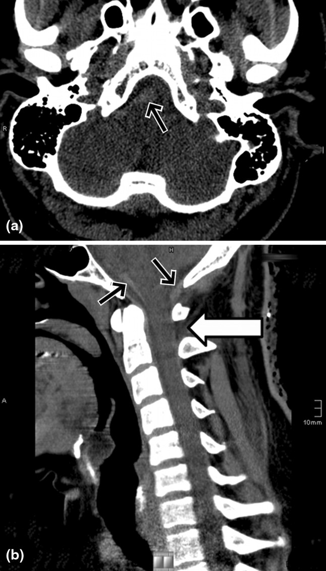
Methods: A 25-year-old man was stabbed at C1/2 and had an incomplete Brown-Sequard syndrome. He underwent surgical exploration because of CSF leakage on the fourth day.

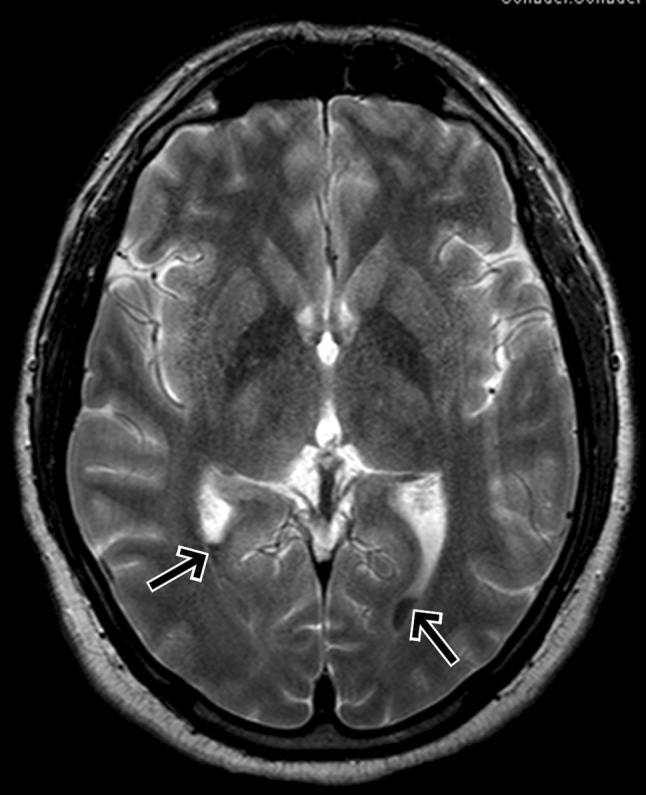
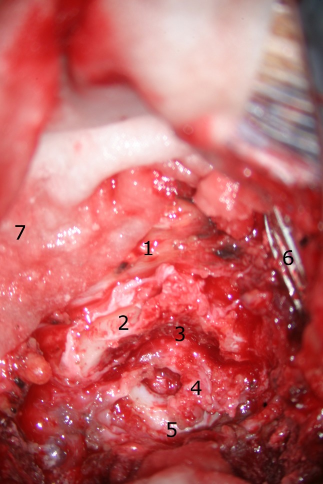
Results: After a follow-up period of 32 months, he was left with a remaining loss of the proprioception of the right foot. We show detailed CT and MR images with the focus on the lesions of the dura and myelon and compared them with intraoperative images. In addition, we contrast our findings with a review of literature published over the last three decades.

Conclusion: MRI gives the most detailed view of soft tissue lesions in SSWs and is in accordance with our intraoperative findings.


Figures

Fig. 1
Fig. 1
a, b Axial CT and reconstructed sagittal MPR showing acute perimedullar hemorrhage (black arrows); a dural defect can hardly be seen (white arrow)
Fig. 2
Fig. 2
MRI axial T2-weighted sequence demonstrating a fluid level in the posterior horn of the ventricles due to hemorrhage
Fig. 3
Fig. 3
MRI T2-weighted sagittal image hypointense structures indicate acute hemorrhage in perimedullar and prepontine location (black arrows) and a laceration of the dorsomedian dura mater between C1/2 (white arrow); a faint hyperintense signal in the posterior parts of the medulla also can be seen
Fig. 4
Fig. 4
MRI T1-weighted sagittal image demonstrating subarachnoid hemorrhage (black arrows) but in this case the dural defect cannot be seen as clearly as in the T2-weighted image
Fig. 5
Fig. 5
MR axial T2-weighted images, showing the absence of the dorsomedian aspect of the hypointense dura (white arrow)
Fig. 6
Fig. 6
MR axial T1-weighted images, showing the absence of the dorsomedian aspect of the hypointense dura (white arrow)
Fig. 7
Fig. 7
The patient is in a prone position and the head fixed with a pinion headholder. The surgeon is at the head of the table, so the caudal direction is at 12 o’clock. The skin is cutted median above the spinous processes and the paravertebral muscles are moved laterally. Overview of the situs: 1 spinous process of C3, 2 right lamina of the axis, 3 area of partial laminectomy, 4 dura lesion, 5 posterior arch of the atlas, 6 retractor, 7 cotton pad
Fig. 8
Fig. 8
Magnification of the dura lesion. On the ground the incision of the myelon is visible. 1 spinous process of C3, 2 right lamina of the axis, 3 area of partial laminectomy, 4 dura lesion, 6 retractor, 8 myelon
Fig. 9
Fig. 9
The myelon lesion is focused for the best view: the cut ends in the myelon. There is no bleeding whether of the myelon nor epi-/subdural anymore. The suction tube is held in the left hand. 4 dura lesion, 8 myelon, 10 cut into the myelon, 11 suction tube
Fig. 10
Fig. 10
Overview of the entire duraplasty before the wound is closed. A partial laminectomy of C1 was necessary to get enough intact dura for tight sutures. 2 right lamina of the axis, 3 area of partial laminectomy, 7 cotton pad, 12 duraplasty, 13 epidural fat, 14 suture

References

    1. Offiah C, Twigg S. Imaging assessment of penetrating craniocerebral and spinal trauma. Clin Radiol. 2009;64:1146–1157. doi: 10.1016/j.crad.2009.06.004. - DOI - PubMed
    1. Burney RE, Maio RF, Maynard F, et al. Incidence, characteristics, and outcome of spinal cord injury at trauma centers in North America. Arch Surg. 1993;128:596–599. doi: 10.1001/archsurg.1993.01420170132021. - DOI - PubMed
    1. Peacock WJ, Shrosbree RD, Key AD. A review of 450 stabwounds of the spinal cord. S Afr Med J. 1977;51:961–964. - PubMed
    1. Waters RL, Sie I, Adkins RH, et al. Motor recovery following spinal cord injury caused by stab wounds: a multicenter study. Paraplegia. 1995;33:98–101. doi: 10.1038/sc.1995.23. - DOI - PubMed
    1. Rubin G, Tallman D, Sagan L, et al. An unusual stab wound of the cervical spinal cord: a case report. Spine. 2001;26:444–447. doi: 10.1097/00007632-200102150-00023. - DOI - PubMed

Publication types