. 2012 May;5(3):375-88.
doi: 10.1242/dmm.007872. Epub 2012 Feb 23.

ROS constitute a convergence nexus in the development of IGF1 resistance and impaired wound healing in a rat model of type 2 diabetes

Affiliations

ROS constitute a convergence nexus in the development of IGF1 resistance and impaired wound healing in a rat model of type 2 diabetes

Milad S Bitar et al. Dis Model Mech. 2012 May.

Abstract

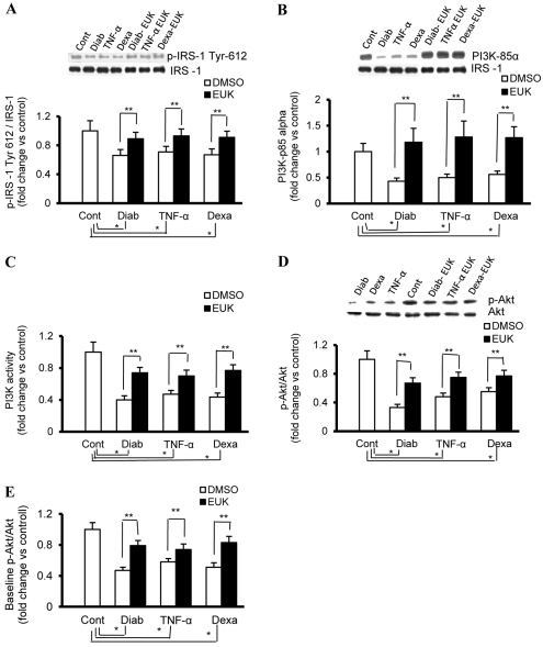
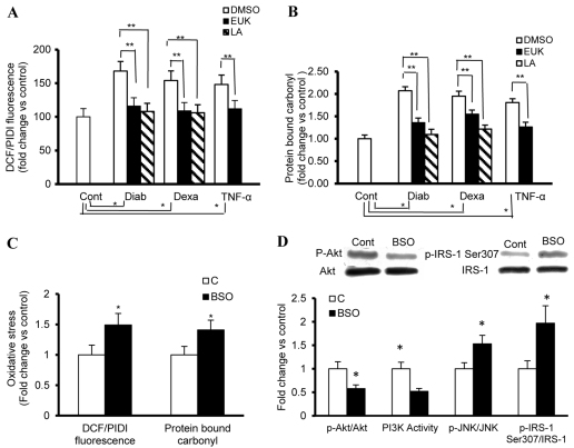
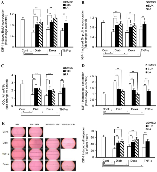
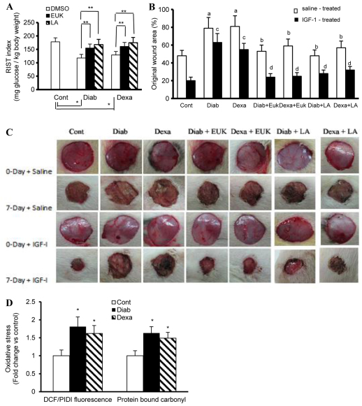
An indolent non-healing wound and insulin and/or insulin-like growth factor (IGF1) resistance are cardinal features of diabetes, inflammation and hypercortisolemia. Little is known about why these phenomena occur in so many contexts. Do the various triggers that induce insulin and/or IGF1 resistance and retard wound healing act through a common mechanism? Cultured dermal fibroblasts from rats and full-thickness excisional wounds were used as models to test the premise that reactive oxygen species (ROS) play a causal role in the development of IGF1 resistance and impaired wound healing under different but pathophysiologically relevant clinical settings, including diabetes, dexamethasone-induced hypercortisolemia and TNFα-induced inflammation. In normal fibroblasts, IGF1 initiated a strong degree of phosphorylation of insulin receptor substrate 1 (IRS1) (Tyr612) and Akt (Ser473), concomitantly with increased PI3K activity. This phenomenon seemed to be attenuated in fibroblasts that had phenotypic features of diabetes, inflammation or hypercortisolemia. Notably, these cells also exhibited an increase in the activity of the ROS-phospho-JNK (p-JNK)-p-IRS1 (Ser307) axis. The above-mentioned defects were reflected functionally by attenuation in IGF1-dependent stimulation of key fibroblast functions, including collagen synthesis and cell proliferation, migration and contraction. The effects of IGF1 on glucose disposal and cutaneous wound healing were also impaired in diabetic or hypercortisolemic rats. The ROS suppressors EUK-134 and α-lipoic acid, or small interfering RNA (siRNA)-mediated silencing of JNK expression, restored IGF1 sensitivity both in vitro and in vivo, and also ameliorated the impairment in IGF1-mediated wound responses during diabetes, inflammation and hypercortisolemia. Our data advance the notion that ROS constitute a convergence nexus for the development of IGF1 resistance and impaired wound healing under different but pathophysiologically relevant clinical settings, with a proof of concept for the beneficial effect of ROS suppressors.


Figures

Fig. 1.
Fig. 1.
Impaired IGF1 signaling in fibroblasts that have phenotypic features of diabetes, inflammation or hypercortisolemia. Fibroblasts with phenotypic characteristics of type 2 diabetes (Diab) and those of control fibroblasts treated chronically with either vehicle (Cont), TNFα (4 ng/ml every day for 4 days) or dexamethasone (Dexa; 20 ng/ml every other day for 8 days) were serum starved for 24 hours prior to treatment for 15 minutes with IGF1 (50 ng/ml). EUK-134 was applied concomitantly with Dexa or TNFα at 100 μM, a concentration that seems to have an optimum therapeutic benefit with a minimum detrimental effect on cell viability. (A,B) Whole-cell protein extracts from IGF1-treated cells were immunoprecipitated with IRS1 and probed with anti-p-IRS1 (Tyr612) (A) or anti-p85α (B) antibody. The membranes were stripped and reprobed with anti-total-IRS1 antibody. (C) PI3K activity in total protein extracts of IGF1-treated cells was determined using an ELISA-based assay (Echelon Biosciences). (D) Phosphorylated (p-Akt-Ser473) and total Akt in protein extracts of IGF1-treated cells were determined using a western-blotting-based technique. (E) Basal expression levels of Akt and its phosphorylated form (p-Akt-Ser473) in paraformaldehyde-fixed cells were quantified using FAC ELISA assays (Active Motif). Experiments were performed in triplicate on each of the cell lines used (n=4 for each). Results represent the means ± s.e.m. *Significantly different from corresponding control values at P≤0.05; **significantly different from corresponding DMSO-treated Diab, TNFα or Dexa values at P≤0.05.
Fig. 2.
Fig. 2.
Genetic inhibition of JNK activity ameliorates the enhancement in p-IRS1 (Ser307) and the defect in PI3K-Akt-dependent signaling during diabetes and hypercortisolemia. (A) Whole-cell protein extracts of control (Cont), diabetic (Diab), dexamethasone (Dexa)- or TNFα-treated fibroblasts were immunoprecipitated with anti-IRS1 and probed with anti-p-IRS (Ser307). The membranes were stripped and reprobed with anti-total-IRS1 antibody. (B) Basal expression levels of JNK and its phosphorylated form (p-JNK) in paraformaldehyde-fixed fibroblasts were quantified using FAC ELISA assays (Active Motif). (C) Cells were transfected with 25 nM si-JNK, as indicated in the Methods, and the level of JNK mRNA was measured using TaqMan real-time PCR. The value of expression relative to that of mock controls (cells treated with transfection reagent only) was normalized against 18S rRNA. (D–F) Fibroblasts with phenotypic features of diabetes or hypercortisolemia were transfected with 25 nM si-JNK 48 hours before exposure to vehicle or IGF1 (50 ng), and cell lysates were collected and used for the assessment of (D) p-IRS1 (Ser307), (E) PI3K activity or (F) p-Akt:Akt ratio as described in the Methods. EUK-134 was applied at 100 μM, a concentration that seems to have an optimum therapeutic benefit with a minimum detrimental effect on cell viability. Experiments were performed in triplicate on each cell line used (n=4 for each). Results represent the means ± s.e.m. *Significantly different from corresponding Cont values at P≤0.05; **significantly different from corresponding DMSO/mock-treated Diab, TNFα or Dexa values at P≤0.05.
Fig. 3.
Fig. 3.
Oxidative stress inhibits IGF-mediated PI3-Akt signaling but activates the JNK–p-IRS1 (Ser307) axis. (A,B) Fibroblasts from a rat model of type 2 diabetes (Diab) and control fibroblasts treated chronically with either vehicle (Cont), TNFα (4 ng/ml every day for 4 days) or dexamethasone (Dexa; 20 ng/ml every other day for 8 days) were used for the evaluation of the status of oxidative stress, as represented by the level of ROS and protein-bound carbonyls. (A) ROS generation of cells cultured in 96-well plates was measured using the fluorescence probes DCF-DA (5 μM). Values are expressed as DCF fluorescence after 1 hour of incubation normalized to total cell number derived by propidium iodide (PI) fluorescence in the presence of 160 μM digitonin (PIDI). (B) Protein-bound carbonyl levels in total cell extracts were determined using an ELISA-based technique. (C,D) Fibroblasts with phenotypic features of HSOS induced by BSO (10 μM) were used for the determination of the level of ROS and protein-bound carbonyls, and key elements of IGF1 signaling. (C) ROS and protein-bound carbonyl levels were measured as described above. (D) Whole-cell protein extracts were used for the assessment of the ratio of p-IRS1 (Ser307):IRS1 (immunoprecipitated with anti-IRS1 and probed with anti-p-IRS1 (Ser307), stripped and reprobed with ant-IRS1), the ratio of p-Akt (Ser473):Akt (western blotting) and PI3K activity (ELISA). In addition, the ratio of p-JNK:JNK in paraformaldehyde-fixed cells was quantified using FAC ELISA assays (Active Motif). EUK-134 or LA was applied at 100 μM (EUK) or 500 μM (LA), concentrations that seem to have an optimum therapeutic benefit with a minimum detrimental effect on cell viability. Experiments were performed in triplicate on each cell lines used (n=4 for each). *Significantly different from corresponding Cont values at P≤0.05; **significantly different from corresponding DMSO-treated Diab, TNFα or Dexa values at P≤0.05.
Fig. 4.
Fig. 4.
Key fibroblast functions that are essential for wound healing are altered as a function of diabetes, low-grade inflammation and hypercortisolemia. Fibroblast from a rat model of type 2 diabetes (Diab) and control fibroblasts treated chronically with either vehicle (Cont), TNFα (4 ng/ml every day for 4 days) or dexamethasone (Dexa; 20 ng/ml every other day for 8 days) were serum starved for 24 hours prior to treatment with IGF1 (50 ng/ml) and used for the assessment of collagen synthesis and cell proliferation, contraction and migration. EUK-134 and LA were used at concentrations of 100 μM and 500 μM, respectively. (A) BrdU incorporation into DNA in cells cultured in 96-well plates was used as an indicator for the determination of the rate of cell proliferation. (B) Radiolabelled proline uptake by cells cultured in 24-well plates was used as an indicator for the measurement of the rate of collagen synthesis. (C) TaqMan real-time PCR conducted on cDNA of cells cultured in six-well plates was used in the determination of the rate of COL1A1 expression. (D) The weight of floating collagen gel matrix of cells cultured in 24-well plates was used for the quantification of the rate of cell contraction. (E) An in vitro scratch wound assay of cells cultured in six-well plates was used to assess the rate of cell migration. Experiments were performed in triplicate for each of the four cell lines (n=4 for each). Results represent the mean ± s.e.m. *Significantly different from corresponding Cont values at P≤0.05; **significantly different from corresponding DMSO-treated Diab, TNFα or Dexa values at P≤0.05.
Fig. 5.
Fig. 5.
Effects of IGF1 on glucose disposal and cutaneous wound healing in control (Cont), diabetic (Diab) and hypercortisolemic (Dexa) animals. (A) IGF1 sensitivity as indicated by the rate of glucose metabolism was evaluated in Cont, Diab (GK rats, a genetic model of type II diabetes) and Dexa (Wistar rats receiving 5 μg Dexa/kg/day, subcutaneously) animals using the RIST with IGF1 (200 μg/kg) infused for 5 minutes instead of insulin. (B,C) Full-thickness excisional wounds induced in Cont, Diab and Dexa animals were subjected to IGF1 and IGFBP1 pluronic acid gel-based therapy. Wound closure was studied at 7 days following wound initiation. (C) Photographs of the macroscopic kinetic analysis of wound sites were taken. (B) Quantitation of data from C, demonstrating the proportion of wound remaining open relative to the initial wound area at the 7-day time point following injury. EUK-134 (12.5 mg/kg) or LA (50 mg/kg) were administered by i.p. injection every other day for 30 days prior to wound induction and this form of therapy continued during the course of wound healing. Similarly, animals used in the IGF1 sensitivity studies were also subjected for 30 days to EUK-134 and LA treatment. (D) Cells isolated from the 7-day diabetic or hypercortisolemic wounds were assessed for oxidative stress using fluorescence- and ELISA-based technique. Results are expressed as the mean ± s.e.m. of six to eight animals/group. *Significantly different from corresponding Cont values at P≤0.05; **significantly different from corresponding DMSO-treated Diab and Dexa animals at P≤0.05; asignificantly different from corresponding Cont saline-treated values at P≤0.05; bsignificantly different from corresponding saline-treated Diab or Dexa values at P≤0.05; csignificantly different from corresponding control IGF1-treated values at P≤0.05; dsignificantly different from corresponding IGF1-treated Diab or Dexa values at P≤0.5.

Similar articles

Cited by

References

    1. Aguirre V., Uchida T., Yenush L., Davis R., White M. F. (2000). The c-Jun NH(2)-terminal kinase promotes insulin resistance during association with insulin receptor substrate-1 and phosphorylation of Ser(307). J. Biol. Chem. 275, 9047–9054 - PubMed
    1. Barja G. (2002). Rate of generation of oxidative stress-related damage and animal longevity. Free Radic. Biol. Med. 33, 1167–1172 - PubMed
    1. Beckert S., Haack S., Hierlemann H., Farrahi F., Mayer P., Konigsrainer A., Coerper S. (2007). Stimulation of steroid-suppressed cutaneous healing by repeated topical application of IGF-I: different mechanisms of action based upon the mode of IGF-I delivery. J. Surg. Res. 139, 217–221 - PubMed
    1. Bennett B. L., Satoh Y., Lewis A. J. (2003). JNK: a new therapeutic target for diabetes. Curr. Opin. Pharmacol. 3, 420–425 - PubMed
    1. Bitar M. S. (1997). Insulin-like growth factor-1 reverses diabetes-induced wound healing impairment in rats. Horm. Metab. Res. 29, 383–386 - PubMed

Publication types

MeSH terms