. 2012 Jul;47(1):104-11.
doi: 10.1165/rcmb.2011-0260OC. Epub 2012 Feb 23.

Role of CXCL5 in leukocyte recruitment to the lungs during secondhand smoke exposure

Affiliations

Role of CXCL5 in leukocyte recruitment to the lungs during secondhand smoke exposure

Gayathriy Balamayooran et al. Am J Respir Cell Mol Biol. 2012 Jul.

Abstract

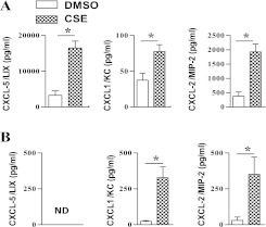
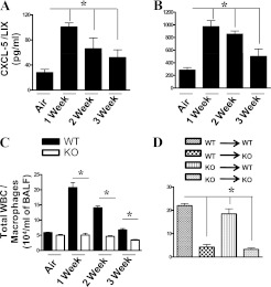
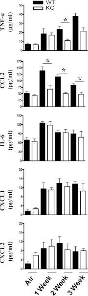
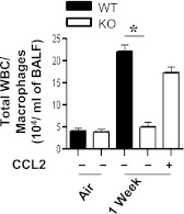
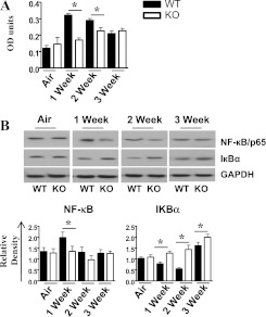
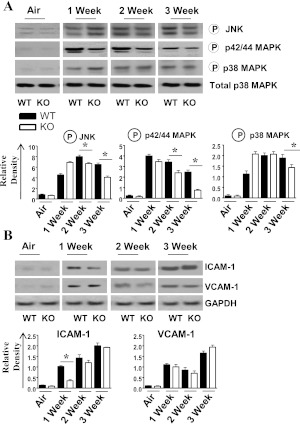
Chronic obstructive pulmonary disease (COPD) is the third leading cause of mortality in the United States. The major cause of COPD is cigarette smoking. Extensive leukocyte influx into the lungs, mediated by chemokines, is a critical event leading to COPD. Although both resident and myeloid cells secrete chemokines in response to inflammatory stimuli, little is known about the role of epithelial-derived chemokines, such as CXC chemokine ligand (CXCL)5, in the pathogenesis of cigarette smoke-induced inflammation. To explore the role of CXCL5, we generated CXCL5 gene-deficient mice and exposed them to secondhand smoke (SHS) for 5 hours/day for 5 days/week up to 3 weeks (subacute exposure). We observed a reduced recruitment of leukocytes to the lungs of CXCL5(-/-) mice compared with their wild-type (WT) counterparts, and noted that macrophages comprised the predominant leukocytes recruited to the lungs. Irradiation experiments performed on CXCL5(-/-) or WT mice transplanted with WT or CXCL5(-/-) bone marrow revealed that resident but not hematopoietic cell-driven CXCL5 is important for mediating SHS-induced lung inflammation. Interestingly, we observed a significant reduction of monocyte chemotactic protein-1 (MCP-1/CC chemokine ligand 2) concentrations in the lungs of CXCL5(-/-) mice. The instillation of recombinant MCP-1 in CXCL5(-/-) mice reversed macrophage recruitment. Our results also show the reduced activation of NF-κB/p65 in the lungs, as well as the attenuated activation of C-Jun N-terminal kinase, p42/44, and p38 mitogen-activated protein kinases and the expression of intercellular adhesion molecule-1 in the lungs of SHS-exposed CXCL5(-/-) mice. Our findings suggest an important role for CXCL5 in augmenting leukocyte recruitment in SHS-induced lung inflammation, and provide novel insights into CXCL5-driven pathogenesis.


Figures

Figure 1.
Figure 1.
Expression of the chemokines CXC chemokine ligand (CXCL)5/lipopolysaccharide-induced CXC chemokine (LIX), CXCL1/keratinocyte-derived chemokine (KC), and CXCL2/macrophage inflammatory protein (MIP)-2 in the culture media of wild-type (WT) alveolar Type II epithelial cells (A) and bone marrow–derived macrophages (B) after cigarette smoke extract (CSE) stimulation (80 μg/ml) in vitro. Experiments were performed in triplicate wells (n = 3–4 mice/group). *P < 0.05, compared with DMSO control. ND, not detected.
Figure 2.
Figure 2.
CXCL5 concentrations in bronchoalveolar lavage fluid (BALF) (A) and lungs (B) after secondhand smoke (SHS) exposure. WT (C57Bl/6) mice were exposed to SHS or HEPA-filtered air, and BALF and lung homogenates were used to detect CXCL5 concentrations by sandwich ELISA (n = 4–5 mice/group; P < 0.05). (C) Total leukocyte/macrophage numbers in the lungs of CXCL5+/+ and CXCL5−/− mice upon cigarette smoke exposure. Mice were exposed to HEPA-filtered air–diluted SHS (10 mg/m3 for 5 hours/day/week) for up to 3 weeks. Control animals were exposed to HEPA-filtered air. At the end of each week, CXCL5−/− and WT mice were killed to determine the number of cells recruited to the lungs, and to perform further analyses as described in Materials and Methods. For cellular enumeration, lungs were lavaged, and the BALF was obtained at 1, 2, and 3 weeks after exposure (n = 8 mice/group; P < 0.05). (D) Total leukocyte/macrophage numbers in the airspaces of bone marrow chimeras of CXCL5+/+ (WT) and CXCL5−/− (knockout; KO) mice after smoke exposure (n = 6–8 mice/group.) WBC, white blood cells. *P < 0.05, compared with CXCL5−/− mice.
Figure 3.
Figure 3.
Cytokine and chemokine concentrations in the lungs after cigarette smoke exposure for up to 3 weeks. Mice were exposed to cigarette smoke, and cell-free BALF was used to determine concentrations (pg/ml) of TNF-α, CCL2, IL-6, CXCL1, and CXCL2 by sandwich ELISA. Asterisks indicate a significant difference between CXCL5+/+ and CXCL5−/− mice (n = 8 mice in each group at each time-point). MCP-1, monocyte chemotactic protein–1. P < 0.05.
Figure 4.
Figure 4.
Effects of exogenous CCL2 on macrophage influx in the lungs of CXCL5−/− mice after SHS exposure. Cellular infiltration was evident in airspaces at 1 week after SHS exposure with MCP-1 (10 μg/mouse) or vehicle (BSA) control. Macrophage influx in BALF from CXCL5−/− or WT mice infected with administration of CCL2 or vehicle (BSA), followed by SHS exposure. For all experiments, n = 5−8 mice/group. *P < 0.05, compared with BSA–administered mice.
Figure 5.
Figure 5.
Activation of NF-κB in whole-lung homogenates after smoke exposure in CXCL5−/− mice. (A) Nuclear lysates from CXCL5−/− mice and control mice were prepared at 1, 2, and 3 weeks after smoke exposure. The NF-κB binding assay was performed in nuclear extracts from the lungs. OD, optical density. (B) Lung homogenates were prepared and total protein from lungs was resolved on an SDS-PAGE, and the membranes were blotted with antibodies against the activated/phosphorylated form of NF-κB and mitogen-activated protein kinases (MAPKs), as described in Materials and Methods. Upper panel in B: Blots are representative of three independent experiments with identical results. Lower panel in B: Relative densities, normalized against glyceraldehyde 3–phosphate dehydrogenase (GAPDH), are representative of three independent blots/experiments. For experiments in both A and B, n = 5–6 mice/group; *P < 0.05, compared with CXCL5−/− mice.
Figure 6.
Figure 6.
(A) Activation of MAPKs in the lungs after SHS exposure in CXCL5−/− mice. Total proteins in the lungs of CXCL5−/− mice and control mice were isolated and resolved on an SDS-PAGE, and the membranes were blotted with the antibodies against activated/phosphorylated or inactive forms of MAPKs, as described in Materials and Methods. Above: Representative of three separate experiments with identical results. Below: Relative densities were normalized against total p38 MAPK, and are representation of three different blots. Circled “P” indicates phosphorylated form of the protein. (B) Expression of intercellular adhesion molecule (ICAM)-1 and vascular cell adhesion molecule (VCAM)-1 in the lung homogenates after subacute smoke exposure. The lungs were homogenized, and total proteins were resolved on SDS-PAGE gel and transferred onto a nitrocellulose membrane. The membranes were blotted with antibodies against ICAM-1, VCAM-1, and GAPDH. This blot is representative of three separate blots with identical results. Below: Densitometric analysis was performed in three blots to demonstrate the expression of ICAM-1 and VCAM-1. For experiments in both panels, n = 4–6 mice/group. *P < 0.05, compared with CXCL5−/− mice.

References

    1. Mannino DM, Buist AS. Global burden of COPD: risk factors, prevalence, and future trends. Lancet 2007;370:765–773 - PubMed
    1. Lopez AD, Murray CCJL. The global burden of disease, 1990–2020. Nat Med 1998;4:1241–1243 - PubMed
    1. Balkissoon R, Lommatzsch S, Carolan B, Make B. Chronic obstructive pulmonary disease: a concise review. Med Clin North Am 2011;95:1125–1141 - PubMed
    1. Barnes PJ, Shapiro SD, Pauwels RA. Chronic obstructive pulmonary disease: molecular and cellular mechanisms. Eur Respir J 2003;22:672–688 - PubMed
    1. Di Stefano A, Caramori G, Ricciardolo FLM, Capelli A, Adcock IM, Donner CF. Cellular and molecular mechanisms in chronic obstructive pulmonary disease: an overview. Clin Exp Allergy 2004;34:1156–1167 - PubMed

Publication types

MeSH terms