. 2012 Apr 6;287(15):12365-78.
doi: 10.1074/jbc.M111.328914. Epub 2012 Feb 23.

A bridging [4Fe-4S] cluster and nucleotide binding are essential for function of the Cfd1-Nbp35 complex as a scaffold in iron-sulfur protein maturation

Affiliations

A bridging [4Fe-4S] cluster and nucleotide binding are essential for function of the Cfd1-Nbp35 complex as a scaffold in iron-sulfur protein maturation

Daili J A Netz et al. J Biol Chem. .

Abstract

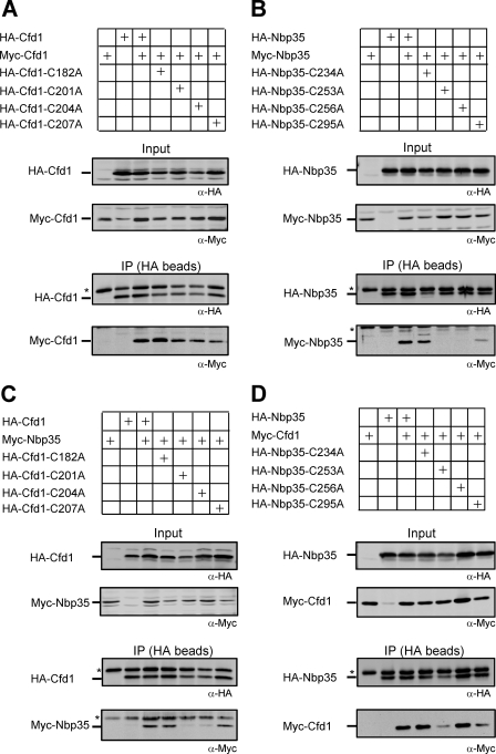
The essential P-loop NTPases Cfd1 and Nbp35 of the cytosolic iron-sulfur (Fe-S) protein assembly machinery perform a scaffold function for Fe-S cluster synthesis. Both proteins contain a nucleotide binding motif of unknown function and a C-terminal motif with four conserved cysteine residues. The latter motif defines the Mrp/Nbp35 subclass of P-loop NTPases and is suspected to be involved in transient Fe-S cluster binding. To elucidate the function of these two motifs, we first created cysteine mutant proteins of Cfd1 and Nbp35 and investigated the consequences of these mutations by genetic, cell biological, biochemical, and spectroscopic approaches. The two central cysteine residues (CPXC) of the C-terminal motif were found to be crucial for cell viability, protein function, coordination of a labile [4Fe-4S] cluster, and Cfd1-Nbp35 hetero-tetramer formation. Surprisingly, the two proximal cysteine residues were dispensable for all these functions, despite their strict evolutionary conservation. Several lines of evidence suggest that the C-terminal CPXC motifs of Cfd1-Nbp35 coordinate a bridging [4Fe-4S] cluster. Upon mutation of the nucleotide binding motifs Fe-S clusters could no longer be assembled on these proteins unless wild-type copies of Cfd1 and Nbp35 were present in trans. This result indicated that Fe-S cluster loading on these scaffold proteins is a nucleotide-dependent step. We propose that the bridging coordination of the C-terminal Fe-S cluster may be ideal for its facile assembly, labile binding, and efficient transfer to target Fe-S apoproteins, a step facilitated by the cytosolic iron-sulfur (Fe-S) protein assembly proteins Nar1 and Cia1 in vivo.


Figures

FIGURE 1.
FIGURE 1.
Effect of cysteine mutagenesis in Cfd1 and Nbp35 on yeast cell growth. The schemes at the top of A and B show the positions of the conserved cysteine residues in Cfd1 and Nbp35, respectively. Plasmids (416) harboring no insert (empty), wild-type (WT), or cysteine mutant versions of CFD1 (A) or NBP35 (B) under control of their respective endogenous promoters were transformed into GalL-CFD1 and GalL-NBP35 yeast cells, respectively. Cells were grown for 40 h in liquid galactose- or glucose-containing minimal medium. 10-Fold serial dilutions were spotted onto agar plates with galactose- or glucose-containing minimal medium. Photographs of the plates were taken after incubation for 48 h at 30 °C.
FIGURE 2.
FIGURE 2.
Identification of Fe-S cluster ligands in Cfd1. A, an empty 416 plasmid or plasmids encoding wild-type (WT) or the indicated mutated HA-tagged Cfd1 proteins under the control of the MET25 promoter were transformed into yeast strain Gal-CFD1. Cells were cultivated in galactose-containing minimal medium for 24 h. Incubation was continued for 16 h in iron-poor minimal medium followed by radiolabeling with 55Fe for 2 h. After preparation of cell extracts with glass beads and affinity isolation of HA-Cfd1, the amounts of Cfd1-bound 55Fe were measured by scintillation counting. B, an empty 416 plasmid or the same plasmid containing untagged WT or cysteine mutant versions of CFD1 under control of the endogenous promoter were transformed into W303 (wild type) or GalL-CFD1 strains. Cells were grown in glucose-containing minimal medium for 40 h, cell extracts were prepared using glass beads, and the isopropyl malate isomerase activity of Leu1 was immediately measured. The lower panels in A and B show immunostains of cell extracts visualizing the amounts of HA-Cfd1 and Leu1, respectively.
FIGURE 3.
FIGURE 3.
Identification of Fe-S cluster ligands in Nbp35. An empty plasmid or plasmids harboring WT or the indicated mutated TAP-tagged (A, 424 plasmids) or HA-tagged (B, 416 plasmids) Nbp35 proteins under the control of the TDH3 (A) or MET25 (B) promoter were transformed into yeast strain Gal-NBP35. Cells were cultivated in glucose-containing minimal medium for 24 h and for another 16 h in iron-poor minimal medium followed by radiolabeling with 55Fe for 2 h. Further analysis for 55Fe incorporation into tagged Nbp35 was according to Fig. 2A. C, an empty 416 plasmid or the same plasmid containing WT or the indicated cysteine mutant versions of NBP35 under control of the own promoter was transformed into W303 (wild-type) or GalL-NBP35 strains. Isopropyl malate isomerase activity was measured as in Fig. 2B. Immunostaining of Nbp35 and Leu1 in cell extracts are presented at the bottom of the panels.
FIGURE 4.
FIGURE 4.
The central two cysteine residues of Cfd1 are required for complex formation with Nbp35. Genes encoding His6-Cfd1 and Nbp35-Strep were cloned into pETDuet-1 multiple cloning sites I and II, respectively. The proteins were overproduced in E. coli and purified by Streptactin affinity chromatography. SDS-PAGE and Coomassie staining of lysates from non-induced (NI) and isopropyl 1-thio-β-d-galactopyranoside-induced cells with Nbp35 and the indicated Cfd1 mutant proteins is shown on the left (asterisks). At the right eluates from Streptactin affinity chromatography of Nbp35 with co-purified wild-type Cfd1 (WT) or indicated Cfd1 mutant proteins are shown. The positions of marker proteins (lane M) and the purified proteins (arrowheads) are indicated.
FIGURE 5.
FIGURE 5.
The two central C-terminal cysteine residues are required for homo- and heteromeric interaction of Cfd1 and Nbp35 in vivo. The indicated wild-type or cysteine mutant versions of Cfd1 (A), Nbp35 (B), or both proteins (C and D) were (co)expressed from centromeric plasmids under control of the MET25 promoter in Gal-CFD1 (A and C) or in Gal-NBP35 (B and D) cells. For Myc- and HA-tagged proteins 414 and 416 plasmids were used, respectively. Cells transformed with the respective plasmids were depleted for Cfd1 or Nbp35 in SC glucose medium for 40 h. Total cell lysates were prepared as described in Fig. 2. Aliquots of the clarified supernatant were TCA-precipitated (Input) or incubated with HA beads for 1 h at 4 °C. After washing, the immunoprecipitated fractions (IP) were analyzed by immunoblotting using monoclonal antibodies against HA (α-HA) and Myc (α-Myc) tags. The asterisks indicate unspecific bands.
FIGURE 6.
FIGURE 6.
Mössbauer spectroscopy of reconstituted Cfd1, Nbp35, and the Cfd1-Nbp35 complex. Mössbauer spectra were recorded at 80 K in the absence of a magnetic field (A–C) or at 4.2 K with a field of 6.5 T applied perpendicular to the γ-rays (D). Open symbols are experimental data points, and colored lines are simulations for component I (red, δ = 0.43 mm s−1, ΔEQ = 1.18 mm s−1, full width at half-maximum (FWHM) = 0.38 mm s−1), component II (blue, δ = 0.34 mm s−1, ΔEQ = 0.62 mm s−1, and FWHM = 0.43 mm s−1), and component III (green, δ = 1.27 mm s−1, ΔEQ = 2.43 mm s−1, and FWHM = 0.55 mm s−1). The black lines correspond to the sum of the three components. The relative intensities of components I, II, and III in the simulations are 54, 34, and 12% for Cfd1, 64, 30, and 6% for Nbp35 and 69, 25, and 6% for the Cfd1-Nbp35 complex, respectively. In D components II and III are broadened beyond detection, and the black line corresponds to a simulation for component I only (δ = 0.45 mm s−1, ΔEQ = 1.19 mm s−1, FWHM = 0.26 mm s−1, and η = 0.70).
FIGURE 7.
FIGURE 7.
No effects of ATP and GTP on the EPR signals from the dithionite-reduced Fe-S clusters of the Cfd1-Nbp35 complex. A, EPR spectra were recorded at a microwave power of 2 milliwatts at 10 K. The buffer contained 4 mm MgCl2 (all cases) and ATP or GTP (2 mm) as indicated. Samples were frozen 2 min after the addition of 4 mm sodium dithionite. B, procedures were as in A but with a microwave power of 20 milliwatt at 4 K (except for the top trace, 10 K). The intense signals at g = 2 have been clipped off. Other EPR parameters were: amplitude modulation, 1 millitesla; microwave frequency, 9.46 GHz; modulation frequency, 100 kHz. C, shown is a rhombogram of the |± 5/2〉 multiplet for a S = 11/2 system. The rhombicity, which gives rise to the sharp isotropic g = 7.26 EPR signal, is highlighted by an arrow.
FIGURE 8.
FIGURE 8.
Mutation of the nucleotide binding motifs of Cfd1 and Nbp35 leads to defective Fe-S cluster loading. A and B, 55Fe incorporation into endogenous Leu1 was followed as described in Fig. 2A for Gal-CFD1 cells (A) and Gal-NBP35 (B) cells grown in galactose- or glucose-containing minimal medium as indicated. C, 55Fe incorporation into plasmid-encoded Cfd1-TAP or its K31A mutant version was measured in Gal-CFD1 cells grown in galactose- or glucose-containing minimal medium as indicated. D, 55Fe incorporation into plasmid-encoded Nbp35-TAP or its K86A mutant version was estimated in Gal-NBP35 cells as described in C. In C and D the background values (around 2 × 10³ cpm/g cells) obtained with empty plasmids were subtracted. Immunostaining of Leu1, Cfd1, and Nbp35 in cell extracts is shown at the bottom of the panels. For all panels 424 plasmids under the control of the TDH3 promoter were used.
FIGURE 9.
FIGURE 9.
Model depicting the two possible geometries for the Cfd1-Nbp35 hetero-tetrameric scaffold complex in the holo form. The N-terminal (N) [4Fe-4S] clusters of Nbp35 and the proposed bridging (B) [4Fe-4S] clusters are indicated. See “Discussion” for further details.

Similar articles

Cited by

References

    1. Beinert H. (2000) Iron-sulfur proteins. Ancient structures, still full of surprises. J. Biol. Inorg. Chem. 5, 2–15 - PubMed
    1. Lill R. (2009) Function and biogenesis iron-sulfur proteins. Nature 460, 831–838 - PubMed
    1. Johnson D. C., Dean D. R., Smith A. D., Johnson M. K. (2005) Structure, function, and formation of biological iron-sulfur clusters. Ann. Rev. Biochem. 74, 247–281 - PubMed
    1. Ayala-Castro C., Saini A., Outten F. W. (2008) Fe-S cluster assembly pathways in bacteria. Microbiol. Mol. Biol. Rev. 72, 110–125 - PMC - PubMed
    1. Lill R., Mühlenhoff U. (2008) Maturation of iron-sulfur proteins in eukaryotes. Mechanisms, connected processes, and diseases. Annu. Rev. Biochem. 77, 669–700 - PubMed

Publication types

MeSH terms