Review
. 2012 May;23(5):774-80.
doi: 10.1681/ASN.2012010029. Epub 2012 Feb 23.

Molecular mechanisms of acid-base sensing by the kidney

Affiliations
Review

Molecular mechanisms of acid-base sensing by the kidney

Dennis Brown et al. J Am Soc Nephrol. 2012 May.

Abstract

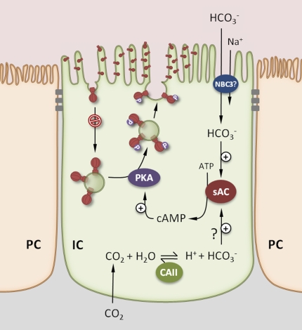
A major function of the kidney is to collaborate with the respiratory system to maintain systemic acid-base status within limits compatible with normal cell and organ function. It achieves this by regulating the excretion and recovery of bicarbonate (mainly in the proximal tubule) and the secretion of buffered protons (mainly in the distal tubule and collecting duct). How proximal tubular cells and distal professional proton transporting (intercalated) cells sense and respond to changes in pH, bicarbonate, and CO(2) status is a question that has intrigued many generations of renal physiologists. Over the past few years, however, some candidate molecular pH sensors have been identified, including acid/alkali-sensing receptors (GPR4, InsR-RR), kinases (Pyk2, ErbB1/2), pH-sensitive ion channels (ASICs, TASK, ROMK), and the bicarbonate-stimulated adenylyl cyclase (sAC). Some acid-sensing mechanisms in other tissues, such as CAII-PDK2L1 in taste buds, might also have similar roles to play in the kidney. Finally, the function of a variety of additional membrane channels and transporters is altered by pH variations both within and outside the cell, and the expression of several metabolic enzymes are altered by acid-base status in parts of the nephron. Thus, it is possible that a master pH sensor will never be identified. Rather, the kidney seems equipped with a battery of molecules that scan the epithelial cell environment to mount a coordinated physiologic response that maintains acid-base homeostasis. This review collates current knowledge on renal acid-base sensing in the context of a whole organ sensing and response process.


Figures

Figure 1.
Figure 1.
The sAC is abundant in kidney intercalated cells (ICs; an A-type acid secreting cell is illustrated here), where it colocalizes extensively with the V-ATPase. sAC generates cAMP in response to an increase in intracellular HCO3, a process that is enhanced by a parallel increase in Ca2+. This cAMP signal results in an accumulation of V-ATPase (shown as ball and stalk-like structures here) at the apical surface of A-ICs, possibly upon phosphorylation (P) of one or more subunits of the V-ATPase. This apical accumulation could occur by increased exocytosis of V-ATPase–containing vesicles, and/or reduced endocytosis of the V-ATPase. The increased [HCO3] in the cell could result from an increase in luminal HCO3 due to, for example, defective proximal tubule reabsorption (e.g., renal Fanconi syndrome) that leads to metabolic acidosis. Activation of ICs by increasing apical V-ATPase is a homeostatic mechanism to correct this acidosis. One proposed apical entry pathway for luminal HCO3 in ICs is the sodium/bicarbonate co-transporter NBC3. Increased intracellular [HCO3] might also result from increased basolateral CO2, which is converted to HCO3 and H+ by cytosolic CAII, which is abundant in ICs. Indeed, an increase in basolateral CO2 stimulates apical proton secretion by A-ICs. A remaining puzzle is how A-type (acid-secreting) and B-type (bicarbonate-secreting) ICs differentially respond to variations in systemic acid/base levels to modulate V-ATPase trafficking into and out of their apical and basolateral membranes, respectively. PC, principal cell.

Similar articles

Cited by

References

    1. Wagner CA, Devuyst O, Bourgeois S, Mohebbi N: Regulated acid-base transport in the collecting duct. Pflugers Arch 458: 137–156, 2009 - PubMed
    1. Wagner CA, Finberg KE, Breton S, Marshansky V, Brown D, Geibel JP: Renal vacuolar H+-ATPase. Physiol Rev 84: 1263–1314, 2004 - PubMed
    1. Schwartz GJ, Al-Awqati Q: Carbon dioxide causes exocytosis of vesicles containing H+ pumps in isolated perfused proximal and collecting tubules. J Clin Invest 75: 1638–1644, 1985 - PMC - PubMed
    1. van Adelsberg J, Al-Awqati Q: Regulation of cell pH by Ca+2-mediated exocytotic insertion of H+-ATPases. J Cell Biol 102: 1638–1645, 1986 - PMC - PubMed
    1. Li S, Sato S, Yang X, Preisig PA, Alpern RJ: Pyk2 activation is integral to acid stimulation of sodium/hydrogen exchanger 3. J Clin Invest 114: 1782–1789, 2004 - PMC - PubMed

Publication types

MeSH terms