. 2013 Feb;62(2):299-309.
doi: 10.1136/gutjnl-2011-301494. Epub 2012 Feb 23.

Hedgehog signalling regulates liver sinusoidal endothelial cell capillarisation

Affiliations

Hedgehog signalling regulates liver sinusoidal endothelial cell capillarisation

Guanhua Xie et al. Gut. 2013 Feb.

Abstract

Objective: Vascular remodelling during liver damage involves loss of healthy liver sinusoidal endothelial cell (LSEC) phenotype via capillarisation. Hedgehog (Hh) signalling regulates vascular development and increases during liver injury. This study therefore examined its role in capillarisation.

Design: Primary LSEC were cultured for 5 days to induce capillarisation. Pharmacological, antibody-mediated and genetic approaches were used to manipulate Hh signalling. Effects on mRNA and protein expression of Hh-regulated genes and capillarisation markers were evaluated by quantitative reverse transcription PCR and immunoblot. Changes in LSEC function were assessed by migration and tube forming assay, and gain/loss of fenestrae was examined by electron microscopy. Mice with acute or chronic liver injury were treated with Hh inhibitors; effects on capillarisation were assessed by immunohistochemistry.

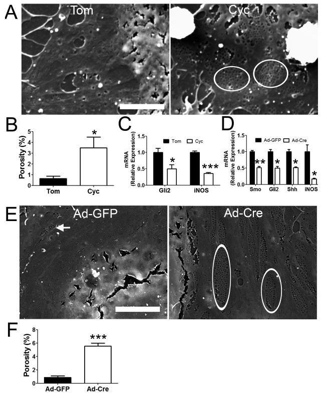
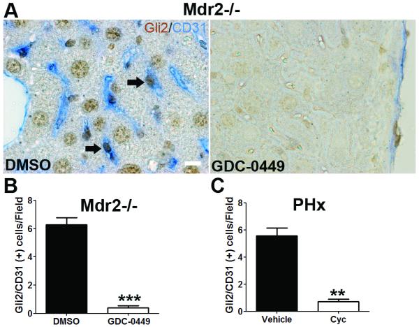
Results: Freshly isolated LSEC expressed Hh ligands, Hh receptors and Hh ligand antagonist Hhip. Capillarisation was accompanied by repression of Hhip and increased expression of Hh-regulated genes. Treatment with Hh agonist further induced expression of Hh ligands and Hh-regulated genes, and upregulated capillarisation-associated genes; whereas Hh signalling antagonist or Hh ligand neutralising antibody each repressed expression of Hh target genes and capillarisation markers. LSEC isolated from Smo(loxP/loxP) transgenic mice that had been infected with adenovirus expressing Cre-recombinase to delete Smoothened showed over 75% knockdown of Smoothened. During culture, Smoothened-deficient LSEC had inhibited Hh signalling, less induction of capillarisation-associated genes and retention of fenestrae. In mice with injured livers, inhibiting Hh signalling prevented capillarisation.

Conclusions: LSEC produce and respond to Hh ligands, and use Hh signalling to regulate complex phenotypic changes that occur during capillarisation.


Figures

Figure 1
Figure 1. Characterization of liver sinusoidal endothelial cells (LSEC) isolated by elutriation method
(A) LSEC cultured for 1 day on collagen coated plate demonstrate typical “cobblestone” morphology under phase contrast microscopy. Scale bar: 100 μm. (B) Scanning electron microscopy shows LSEC have fenestrae grouped into sieve plates (arrow), the defining morphology of LSEC. Scale bar: 5 μm. (C) FACS analysis of freshly isolated LSEC. ~96% of LSEC were double positive for CD31 and CD105(endoglin). Only 2% of LSEC were F4/80 positive (Kupffer cell marker) and none expressed desmin (hepatic stellate cell marker). Uptake of Ac-Di-LDL (D) and FITC-FSA (E) by day 1 LSEC demonstrate the typical endocytic activity of the cells. Scale bar: 50 μm for left images, 20 μm for right images. All experiments were performed at least three times.
Figure 2
Figure 2. LSEC undergo capillarisation spontaneously in vitro
(A) Rat LSEC were cultured on collagen coated plates, processed and examined by SEM at X10,000 magnification. Day 1 LSEC show numerous fenestrae grouped into sieve plates (arrow). Day 2 and 3 LSEC have fewer fenestrae and sieve plates (arrow). Day 4 and 5 LSEC have only occasional fenestrae (arrowhead). Porosity measurement was used to quantify the percentage of fenestrae per surface area. Scale bar: 5 μm. **: p<0.01 day 1 cells, n=3. (B) qRT-PCR analysis of endothelial cell associated gene expression changes during culture induced capillarisation. * p<0.05, ** p<0.01, *** p<0.001 vs day 0 cells, n=3. (C) Western blot analysis of protein harvested from rat LSEC with densitometry to confirm the gene expression change described in B. Results are representative of triplicate experiments. * p<0.05, ** p<0.01 vs day 0 cells, # non-detectable.
Figure 3
Figure 3. Hedgehog (Hh) pathway is activated during LSEC capillarisation in vitro
(A) Rat LSEC were cultured, mRNA were harvested and qRT-PCR were used to analysis the expression change of Hh target genes (Gli2, Gli3, Sonic Hh (Shh), Hedgehog-interacting protein (Hhip), Patched (Ptc), Smoothened, Twist2, secreted frizzled-related protein 1 (sFRP1), osteopontin (OPN)). * p<0.05, ** p<0.01, *** p<0.001 vs day 0 cells, n=3. (B) Western blot analysis of protein harvested from rat LSEC with densitometry to confirm the gene expression change described in B. β-Actin was used as loading control. Results are representative of triplicate experiments. * p<0.05, ** p<0.01, *** p<0.001 vs day 0 cells, # non-detectable.
Figure 4
Figure 4. LSEC are Hh responsive
(A) Rat LSEC were cultured for 3 days and treated with either DMSO or SAG (0.3 μM, an Hh agonist) for 2 more days. mRNA were isolated and gene expression changes were examined by qRT-PCR. (B) Day 3 rat LSEC were treated with either cyclopamine (Cyc, 3μM, a pharmacological inhibitor of Hh signaling) or tomatidine (Tom, an inert cyclopamine analog) for 2 days. mRNA was harvested, and changes in gene expression were determined by qRT-PCR. (C) Rat LSEC were cultured for 3 days and treated with either 5E1(10 μg/ml, Hh ligand neutralizing antibody) or IgG for 2 more days. mRNA was isolated and gene expression change were monitored by qRT-PCR. *: p<0.05, **: p<0.01, ***: p<0.001. n= 3.
Figure 5
Figure 5. Effect of knocking down LSEC Smo gene expression in vitro on Hh pathway
(A) Primary LSEC were isolated from SmoloxP/loxP transgenic mice, placed in monocultures for 3 days, and infected with either adenovirus expressing GFP (Ad-GFP, control) or Cre-recombinase (Ad-Cre, for conditional deletion of the Hh signaling intermediate, Smo). After 48 h, cells were harvested to obtain mRNA for qRT-PCR analysis. *:p<0.05, **:p<0.01, ***:p<0.001 vs control. n =3. (B) Western blot analysis of protein harvested from mouse LSEC described in A with densitometry to confirm the gene expression change. β-Actin was used as loading control. *:p<0.05, **:p<0.01 vs control. Results are representative of triplicate experiments.
Figure 6
Figure 6. Hh pathway promotes LSEC migration in vitro
Primary rat LSEC were seeded on collagen coated insert and treated with (A) normal growth medium (ctrl), (B) 40ng/ml VEGF, (C) 3μM Cyclopamine (Cyc), (D) VEGF with Cyclopamine (VEGF + Cyc), or (E) 0.3 μM SAG for 20 hours. (F) Effects on migration were assessed by counting the number of cells in the bottom of the inserts in triplicate experiments. Representative micrographs and statistical summary are shown. *: p<0.05, **: p<0.01 vs control. #: p<0.05 vs VEGF. Scale bar: 50 μm.
Figure 7
Figure 7. Hh pathway promotes LSEC tube formation in vitro
Primary rat LSEC were seeded on growth factor reduced Matrigel and treated with (A) normal growth medium (ctrl), (B) 40ng/ml VEGF, (C) 3μM Cyclopamine (Cyc), (D) VEGF with Cyclopamine (VEGF + Cyc), or (E) 0.3 μM SAG for 6 hours. (F) Effects on vascular tube formation were assessed by quantifying the length of capillary like-tube in triplicate experiments. Representative micrographs and statistical summary are shown. *: p<0.05 vs control. #: p<0.05 vs VEGF. Scale bar: 100 μm.
Figure 8
Figure 8. Inhibition of Hh pathway prevents LSEC capillarisation in vitro
Primary rat LSEC were plated on collagen coated coverslip. After 3 hours, LSEC attached and were then incubated with either tomatidine (Tom) or cyclopamine (Cyc, 3μM). Cells were fixed and processed for SEM on day 2. Representative (A) SEM micrograph and (B) porosity measurement were shown. LSEC treated with cyclopamine maintained fenestrae grouped into sieve plates (circle), while LSEC treated with tomatidine lose fenestrae. (C) mRNA were also isolated from the treated cells described in (A) for qRT-PCR analysis on Gli2 and iNOS gene expression. SmoloxP/loxP transgenic mice were pretreated with adenovirus expressing GFP (Ad-GFP) or Cre (Ad-Cre) by tail vein injection. Primary LSEC were isolated 2 days after treatment and plated on collagen-coated dish for another 2 days. (D) mRNA expression of Smo, Gli2, Shh and iNOS were examined by qRT-PCR. Cells were also fixed for (E) SEM micrograph and (F) porosity measurement was shown. Ad-Cre infected LSEC maintain normal fenestrae grouped into sieve plates (circle), while Ad-GFP infected LSEC lose fenestrae. Note that occasional fenestrae and sieve plates (arrow) can still be observed in Ad-GFP infected LSEC, indicating that the loss of fenestrae was not due to the contamination of other kinds of endothelial cells. *: p<0.05, **:p<0.01, ***p<0.001. n=3-6. Scale bar: 5 μm.
Figure 9
Figure 9. Inhibition of Hh pathway prevents LSEC capillarisation in vivo
(A) Liver sections from DMSO and GDC-0449 treated Mdr2−/− mice were double stained for Gli2 (brown, Hh target gene) and CD31 (blue, capillarisation marker). Note that LSEC co-express Gli2 and CD31 (arrow). Scale bar: 10μm. Number of Gli2/CD31 double positive cells per field (B) was counted in 5 random fields per mice, ***p<0.001. n=3. (C) Liver sections from vehicle and cyclopamine treated PHx mice were stained for Gli2 and CD31, and the number of Gli2/CD31 double positive cells was counted. **p<0.01. n=3.

References

    1. Schaffner F, Poper H. Capillarisation of hepatic sinusoids in man. Gastroenterology. 1963;44:239–42. - PubMed
    1. DeLeve LD, Wang X, Kanel GC, et al. Prevention of hepatic fibrosis in a murine model of metabolic syndrome with nonalcoholic steatohepatitis. Am J Pathol. 2008;173:993–1001. - PMC - PubMed
    1. Xu B, Broome U, Uzunel M, et al. Capillarisation of hepatic sinusoid by liver endothelial cell-reactive autoantibodies in patients with cirrhosis and chronic hepatitis. Am J Pathol. 2003;163:1275–89. - PMC - PubMed
    1. Warren A, Bertolino P, Benseler V, et al. Marked changes of the hepatic sinusoid in a transgenic mouse model of acute immune-mediated hepatitis. J Hepatol. 2007;46:239–46. - PubMed
    1. Horn T, Junge J, Christoffersen P. Early alcoholic liver injury: changes of the Disse space in acinar zone 3. Liver. 1985;5:301–10. - PubMed

Publication types

MeSH terms