. 2012 Feb 24;335(6071):970-3.
doi: 10.1126/science.1215156.

Control of nonapoptotic developmental cell death in Caenorhabditis elegans by a polyglutamine-repeat protein

Affiliations

Control of nonapoptotic developmental cell death in Caenorhabditis elegans by a polyglutamine-repeat protein

Elyse S Blum et al. Science. .

Abstract

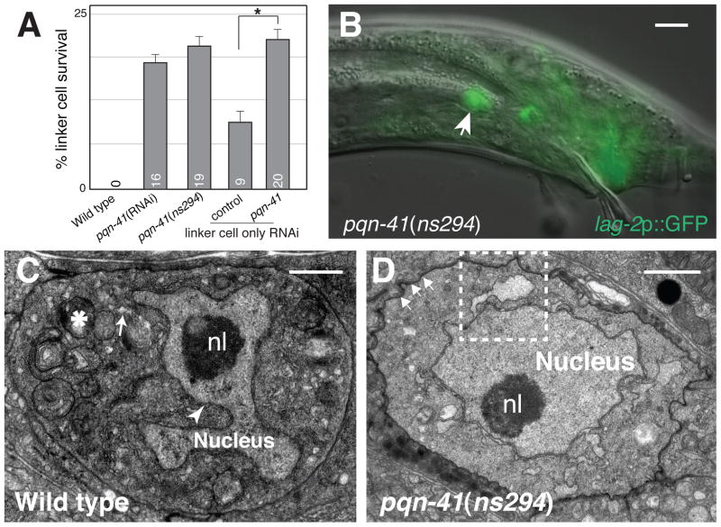
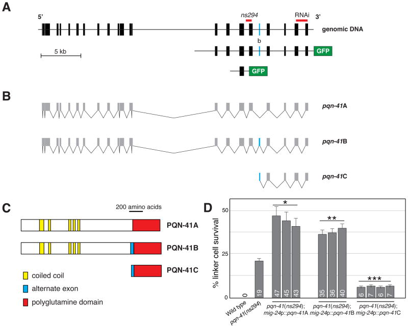
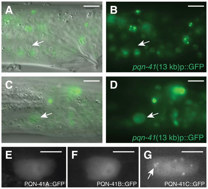
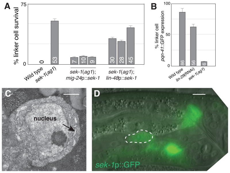
Death is a vital developmental cell fate. In Caenorhabditis elegans, programmed death of the linker cell, which leads gonadal elongation, proceeds independently of caspases and apoptotic effectors. To identify genes promoting linker-cell death, we performed a genome-wide RNA interference screen. We show that linker-cell death requires the gene pqn-41, encoding an endogenous polyglutamine-repeat protein. pqn-41 functions cell-autonomously and is expressed at the onset of linker-cell death. pqn-41 expression is controlled by the mitogen-activated protein kinase kinase SEK-1, which functions in parallel to the zinc-finger protein LIN-29 to promote cellular demise. Linker-cell death is morphologically similar to cell death associated with normal vertebrate development and polyglutamine-induced neurodegeneration. Our results may therefore provide molecular inroads to understanding nonapoptotic cell death in metazoan development and disease.


Figures

Fig. 1
Fig. 1
PQN-41 is required for linker cell death. (A) Linker cell survival 2–4 hours after the L4 molt. Numbers, percentages. Error bars, SEM. Asterisk, p<0.04. n≥50. Increased survival in rde-1 may reflect reduced let-7 miRNA function (8). (B) Adult pqn-41(ns294) male with surviving linker cell (arrow). Scale bar, 10 μm. (C) Electron micrograph of dying linker cell in a wild-type animal. nl, nucleolus. Swollen ER and mitochondria, arrow, asterisk. Arrowhead, nuclear envelope crenellation. (D) Electron micrograph of linker cell in (B). Arrows, adhesion junctions. Dashed box, enlarged in fig. S2A. Scale bars in (C, D), 2 μm.
Fig. 2
Fig. 2
PQN-41C promotes linker cell death. (A) pqn-41 genomic region. Alternative exon b is labeled with the letter “b”. GFP not to scale. (B) Three mRNAs generated by the pqn-41 region. (C) Predicted protein structures for mRNAs in (B). (D) Linker cell survival in pqn-41(ns294) mutants expressing the indicated cDNAs. Numbers, percentages. Error bars, SEM. *, different from pqn-41(ns294), p<0.0009, n=138. **, p<0.0007, n=236. ***, p<0.004, n=229.
Fig. 3
Fig. 3
PQN-41 is expressed as the linker cell dies. (A) DIC and fluorescence image of late L4 male expressing the 13 kb pqn-41::GFP reporter in Fig. 2A. Arrow, linker cell. (B) Fluorescence image only. (C) Same as (A) except in an older L4 animal. (D) Fluorescence image only. (E–G) Expression of mig-24 promoter::pqn-41A, B, and C cDNA GFP translational fusions, respectively. Arrow, cytoplasmic puncta. Scale bars, 10 μm (A–D), 5 μm (E–G).
Fig. 4
Fig. 4
SEK-1 MAPKK is required for linker cell death. (A) Linker cell survival in sek-1(ag1) mutants expressing the indicated promoter::cDNA constructs. Numbers, percentages. Error bars, SEM. n≥70. (B) Expression of the 2.5 kb pqn-41::GFP reporter in late L4 (wild-type) or young adult (lin-29, sek-1) animals. n≥50. (C) EM of linker cell in fig. S9A. Arrow, swollen ER. Scale bar, 2 μm. (D) Late L4 male expressing a sek-1 promoter::GFP reporter. Scale bar, 5 μm.

Comment in

Similar articles

Cited by

References

    1. Abraham MC, Shaham S. Trends Cell Biol. 2004;14:184. - PubMed
    1. Fuchs Y, Steller H. Cell. 2011;147:742. - PMC - PubMed
    1. Kerr JF, Wyllie AH, Currie AR. Br J Cancer. 1972;26:239. - PMC - PubMed
    1. Honarpour N, et al. Dev Biol. 2000;218:248. - PubMed
    1. Coucouvanis E, Martin GR. Cell. 1995;83:279. - PubMed

Publication types

MeSH terms