

Histone deacetylase complexes promote trinucleotide repeat expansions
- PMID: 22363205
- PMCID: PMC3283555
- DOI: 10.1371/journal.pbio.1001257
Histone deacetylase complexes promote trinucleotide repeat expansions
Abstract
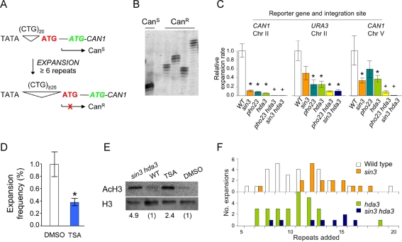
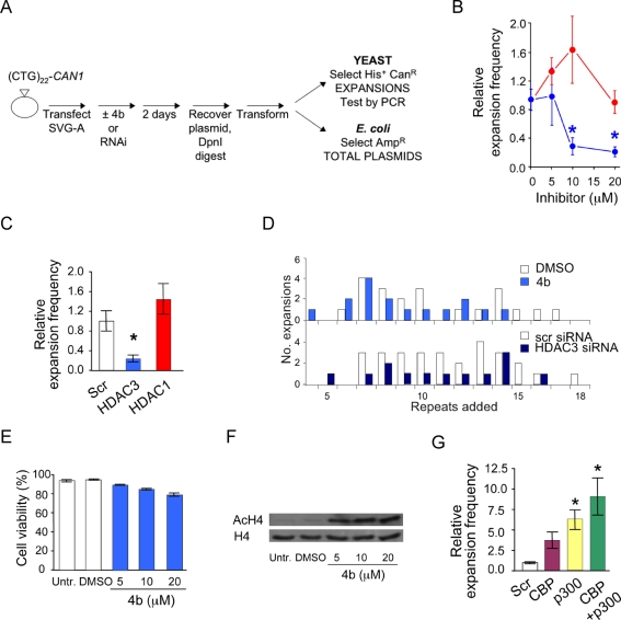
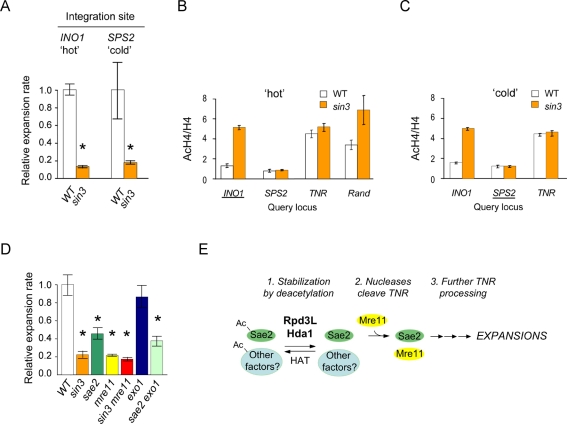
Expansions of DNA trinucleotide repeats cause at least 17 inherited neurodegenerative diseases, such as Huntington's disease. Expansions can occur at frequencies approaching 100% in affected families and in transgenic mice, suggesting that specific cellular proteins actively promote (favor) expansions. The inference is that expansions arise due to the presence of these promoting proteins, not their absence, and that interfering with these proteins can suppress expansions. The goal of this study was to identify novel factors that promote expansions. We discovered that specific histone deacetylase complexes (HDACs) promote CTG•CAG repeat expansions in budding yeast and human cells. Mutation or inhibition of yeast Rpd3L or Hda1 suppressed up to 90% of expansions. In cultured human astrocytes, expansions were suppressed by 75% upon inhibition or knockdown of HDAC3, whereas siRNA against the histone acetyltransferases CBP/p300 stimulated expansions. Genetic and molecular analysis both indicated that HDACs act at a distance from the triplet repeat to promote expansions. Expansion assays with nuclease mutants indicated that Sae2 is one of the relevant factors regulated by Rpd3L and Hda1. The causal relationship between HDACs and expansions indicates that HDACs can promote mutagenesis at some DNA sequences. This relationship further implies that HDAC3 inhibitors being tested for relief of expansion-associated gene silencing may also suppress somatic expansions that contribute to disease progression.
Conflict of interest statement
The authors have declared that no competing interests exist.
Figures
Comment in
-
Enzyme inhibitor may offer dual protection against brain disease.PLoS Biol. 2012 Feb;10(2):e1001270. doi: 10.1371/journal.pbio.1001270. Epub 2012 Feb 21. PLoS Biol. 2012. PMID: 22363209 Free PMC article.
Similar articles
-
MutSβ and histone deacetylase complexes promote expansions of trinucleotide repeats in human cells.Nucleic Acids Res. 2012 Nov 1;40(20):10324-33. doi: 10.1093/nar/gks810. Epub 2012 Aug 31. Nucleic Acids Res. 2012. PMID: 22941650 Free PMC article.
-
HDAC3 deacetylates the DNA mismatch repair factor MutSβ to stimulate triplet repeat expansions.Proc Natl Acad Sci U S A. 2020 Sep 22;117(38):23597-23605. doi: 10.1073/pnas.2013223117. Epub 2020 Sep 8. Proc Natl Acad Sci U S A. 2020. PMID: 32900932 Free PMC article.
-
The Saccharomyces cerevisiae Mre11-Rad50-Xrs2 complex promotes trinucleotide repeat expansions independently of homologous recombination.DNA Repair (Amst). 2016 Jul;43:1-8. doi: 10.1016/j.dnarep.2016.04.012. Epub 2016 May 2. DNA Repair (Amst). 2016. PMID: 27173583
-
Histone deacetylase complexes as caretakers of genome stability.Epigenetics. 2012 Aug;7(8):806-10. doi: 10.4161/epi.20922. Epub 2012 Jun 22. Epigenetics. 2012. PMID: 22722985 Free PMC article. Review.
-
Disease-associated repeat instability and mismatch repair.DNA Repair (Amst). 2016 Feb;38:117-126. doi: 10.1016/j.dnarep.2015.11.008. Epub 2015 Dec 12. DNA Repair (Amst). 2016. PMID: 26774442 Review.
Cited by
-
DNA triplet repeat expansion and mismatch repair.Annu Rev Biochem. 2015;84:199-226. doi: 10.1146/annurev-biochem-060614-034010. Epub 2015 Jan 2. Annu Rev Biochem. 2015. PMID: 25580529 Free PMC article. Review.
-
The Chromatin Remodeler Isw1 Prevents CAG Repeat Expansions During Transcription in Saccharomyces cerevisiae.Genetics. 2018 Mar;208(3):963-976. doi: 10.1534/genetics.117.300529. Epub 2018 Jan 5. Genetics. 2018. PMID: 29305386 Free PMC article.
-
The role of genetics in the establishment and maintenance of the epigenome.Cell Mol Life Sci. 2013 May;70(9):1543-73. doi: 10.1007/s00018-013-1296-2. Epub 2013 Mar 10. Cell Mol Life Sci. 2013. PMID: 23474979 Free PMC article. Review.
-
Expanded complexity of unstable repeat diseases.Biofactors. 2013 Mar-Apr;39(2):164-75. doi: 10.1002/biof.1060. Epub 2012 Dec 11. Biofactors. 2013. PMID: 23233240 Free PMC article. Review.
-
Histone deacetylase-3 interacts with ataxin-7 and is altered in a spinocerebellar ataxia type 7 mouse model.Mol Neurodegener. 2013 Oct 27;8:42. doi: 10.1186/1750-1326-8-42. Mol Neurodegener. 2013. PMID: 24160175 Free PMC article.
References
-
- Orr H. T, Zoghbi H. Y. Trinucleotide repeat disorders. Annu Rev Neurosci. 2007;30:575–621. - PubMed
-
- Mirkin S. M. Expandable DNA repeats and human disease. Nature. 2007;447:932–940. - PubMed
-
- Castel A. L, Cleary J. D, Pearson C. E. Repeat instability as the basis for human diseases and as a potential target for therapy. Nat Rev Mol Cell Biol. 2010;11:165–170. - PubMed
-
- Fu Y-H, Kuhl D. P. A, Pizzuti A, Pieretti M, Sutcliffe J. S, et al. Variation of the CGG repeat at the fragile X site results in genetic instability: resolution of the Sherman paradox. Cell. 1991;67:1047–1058. - PubMed
Publication types
MeSH terms
Substances
LinkOut - more resources
Full Text Sources
Molecular Biology Databases
Miscellaneous
