. 2012 Feb 21:6:26.
doi: 10.3389/fnins.2012.00026. eCollection 2012.

Sexual differentiation of the rodent brain: dogma and beyond

Affiliations

Sexual differentiation of the rodent brain: dogma and beyond

Kathryn M Lenz et al. Front Neurosci. .

Abstract

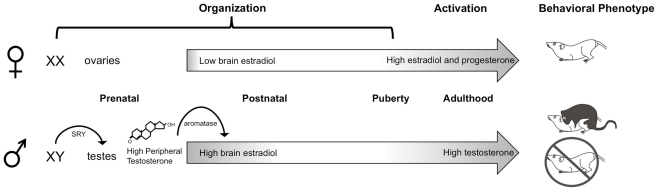
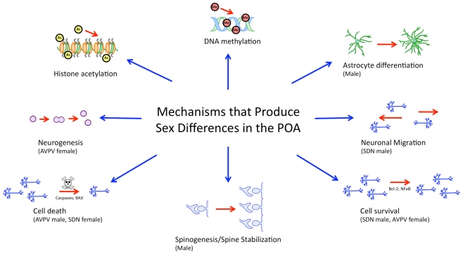
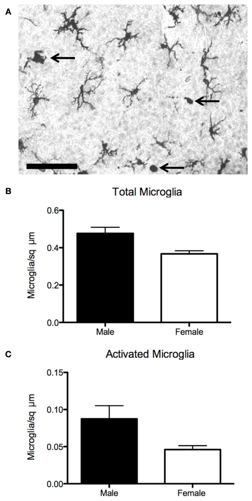
Steroid hormones of gonadal origin act on the neonatal brain to produce sex differences that underlie adult reproductive physiology and behavior. Neuronal sex differences occur on a variety of levels, including differences in regional volume and/or cell number, morphology, physiology, molecular signaling, and gene expression. In the rodent, many of these sex differences are determined by steroid hormones, particularly estradiol, and are established by diverse downstream effects. One brain region that is potently organized by estradiol is the preoptic area (POA), a region critically involved in many behaviors that show sex differences, including copulatory and maternal behaviors. This review focuses on the POA as a case study exemplifying the depth and breadth of our knowledge as well as the gaps in understanding the mechanisms through which gonadal hormones produce lasting neural and behavioral sex differences. In the POA, multiple cell types, including neurons, astrocytes, and microglia are masculinized by estradiol. Multiple downstream molecular mediators are involved, including prostaglandins, various glutamate receptors, protein kinase A, and several immune signaling molecules. Moreover, emerging evidence indicates epigenetic mechanisms maintain sex differences in the POA that are organized perinatally and thereby produce permanent behavioral changes. We also review emerging strategies to better elucidate the mechanisms through which genetics and epigenetics contribute to brain and behavioral sex differences.

Keywords: development; epigenetics; estradiol; hormone; preoptic area; sex difference.


Figures

Figure 1
Figure 1
Neonatal organization of the brain and behavior. In male rodents, the SRY gene on the Y chromosome leads to the development of testes, which begin synthesizing androgens prenatally. Plasma testosterone levels rapidly increase late during the embryonic period and peak shortly after birth. Once in the brain, testosterone is converted by the p450 aromatase enzyme into estradiol, which acts to masculinize and defeminize the brain. A second organizational period occurs peripubertally, when steroid hormones begin to rise. In adulthood, the differentiated brain is activated by high levels of circulating gonadal hormones: androgens in males and estrogens and progestins in females, to produce sex differences in copulatory behaviors.
Figure 2
Figure 2
Mechanisms governing sexual differentiation of the preoptic area. Sexual differentiation of the preoptic area results from the complex interplay of diverse hormonally regulated mechanisms, including neurogenesis, cell survival and death, morphological differentiation of individual cells, and epigenetic activity. These mechanisms are subregion specific, cell-type specific, and occur during temporally limited critical periods during development.
Figure 3
Figure 3
Sex differences in microglia in the preoptic area. (A) Established sex differences in number and morphology of neurons and astrocytes in the preoptic are being joined by sex differences in microglia, the brain’s resident immune cell. (B) Males have more microglia than females on postnatal day 2, when stained for the microglial marker Iba1. (C) Males also have more activated, ameboid microglia (arrows in 3A), which are associated with secretion of prostanoids, cytokines, chemokines, as well as phagocytosis. Scale bar = 50 μm.

References

    1. Abraham I. M., Todman M. G., Korach K. S., Herbison A. E. (2004). Critical in vivo roles for classical estrogen receptors in rapid estrogen actions on intracellular signaling in mouse brain. Endocrinology 145, 3055–306110.1210/en.2003-1676 - DOI - PubMed
    1. Adachi S., Fujioka H., Kakehashi C., Matsuwaki T., Nishihara M., Akema T. (2009). Possible involvement of microglia containing cyclooxygenase-1 in the accumulation of gonadotrophin-releasing hormone in the preoptic area in female rats. J. Neuroendocrinol. 21, 1029–103710.1111/j.1365-2826.2009.01928.x - DOI - PubMed
    1. Ahmed E. I., Zehr J. L., Schulz K. M., Lorenz B. H., DonCarlos L. L., Sisk C. L. (2008). Pubertal hormones modulate the addition of new cells to sexually dimorphic brain regions. Nat. Neurosci. 11, 995–99710.1038/nn.2178 - DOI - PMC - PubMed
    1. Alboni S., Cervia D., Sugama S., Conti B. (2010). Interleukin 18 in the CNS. J. Neuroinflammation 7.10.1186/1742-2094-7-9 - DOI - PMC - PubMed
    1. Amateau S. K., McCarthy M. M. (2002a). A novel mechanism of dendritic spine plasticity involving estradiol induction of prostaglandin-E2. J. Neurosci. 22, 8586–8596 - PMC - PubMed

LinkOut - more resources