. 2012 Feb 20:6:5.
doi: 10.3389/fncir.2012.00005. eCollection 2012.

Possible role of acetylcholine in regulating spatial novelty effects on theta rhythm and grid cells

Affiliations

Possible role of acetylcholine in regulating spatial novelty effects on theta rhythm and grid cells

Caswell Barry et al. Front Neural Circuits. .

Abstract

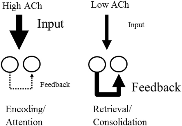
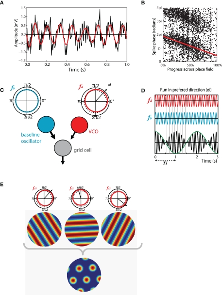
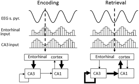
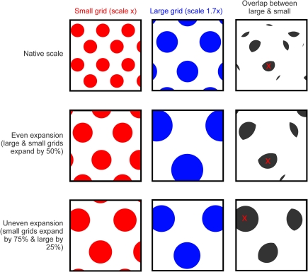
Existing pharmacological and lesion data indicate that acetylcholine plays an important role in memory formation. For example, increased levels of acetylcholine in the hippocampal formation are known to be associated with successful encoding while disruption of the cholinergic system leads to impairments on a range of mnemonic tasks. However, cholinergic signaling from the medial septum also plays a central role in generating and pacing theta-band oscillations throughout the hippocampal formation. Recent experimental results suggest a potential link between these distinct phenomena. Environmental novelty, a condition associated with strong cholinergic drive, has been shown to induce an expansion in the firing pattern of entorhinal grid cells and a reduction in the frequency of theta measured from the LFP. Computational modeling suggests the spatial activity of grid cells is produced by interference between neuronal oscillators; scale being determined by theta-band oscillations impinging on entorhinal stellate cells, the frequency of which is modulated by acetylcholine. Here we propose that increased cholinergic signaling in response to environmental novelty triggers grid expansion by reducing the frequency of the oscillations. Furthermore, we argue that cholinergic induced grid expansion may enhance, or even induce, encoding by producing a mismatch between expanded grid cells and other spatial inputs to the hippocampus, such as boundary vector cells. Indeed, a further source of mismatch is likely to occur between grid cells of different native scales which may expand by different relative amounts.

Keywords: acetylcholine; grid cell; place cell; stellate cell; theta.


Figures

Figure 1
Figure 1
Effect of acetylcholine on cortical processing. High acetylcholine (ACh) enhances encoding by increasing the efficacy of afferent input to the cortex via the action of nicotinic receptors. At the same time high acetylcholine suppresses transmission at glutamatergic recurrent feedback synapses within cortical regions. Conversely, lower acetylcholine favors the retrieval of stored memories by enhancing the influence of recurrent connections relative to afferent input.
Figure 2
Figure 2
Theta-band oscillations play an important role in structuring spatial activity in the hippocampus and entorhinal cortex. (A) Theta recorded from the entorhinal LFP of a moving rat. Black trace, raw LFP, mean normalized showing 1s of data. 8 Hz theta modulation is clearly visible, higher frequency gamma oscillations are also present. Red trace, same signal band pass filtered in 6–12 Hz range. (B) Phase precession in a CA1 place cell, the phase, and location of individual spikes is shown as follows: X-axis indicates the animal's progress across the place field, moving left to right. Y-axis indicates the phase of each spike relative to theta measured from the cell layer, two cycles are shown for completeness. Red line indicates best fit to data for linear-circular regression. (C) Schematic of the basic oscillatory interference model showing two components: a baseline oscillation (blue) with a constant frequency in the theta-band (fb) and; A velocity controlled component (red) the frequency (fd) of which increases proportionate to the animal's velocity in a preferred direction (φ i). (D) Interference pattern generated between the components described in C. Spikes are emitted at the peaks of the carrier (black) which has a frequency equal to the mean of fb and fd. Grid scale is determined by the envelope (green) which has a frequency equal to the difference between fb and fd. (E) Multiple velocity controlled components with preferred firing directions at increments of 60° are required to produce a grid interference pattern. A single baseline oscillator and velocity controlled component will produce a spatially stable stripped pattern similar to a sine grating.
Figure 3
Figure 3
Theta phase is proposed to segment retrieval and encoding phases of mnemonic processing in the hippocampus. Encoding (left) occurs around the peak of theta (measured from the cell-layer) when strong current sinks are evident in the lacunosum-moleculare where entorhinal inputs terminate. Retrieval (right) occurs during the trough of theta when internal connectivity within the hippocampus is more active, producing a strong current sink in the stratum radiatum.
Figure 4
Figure 4
Grid expansion as a source of mismatch. Top row, cartoon showing the spatial activity of two idealized grid cells in a square environment, a small-scale grid is shown in red and larger scale grid (1.7x larger than the small grid) in blue. Panel to the right shows areas where the firing of the two grids overlap (gray), the red cross indicates the area of largest overlap. Middle row, grid activity from the first row with a 50% expansion applied to both the small and large grid (expansion focused on the center of the environment). Fields from both grids are translocated relative to the environment and will likely “mismatch” with spatial inputs, such as boundary vector cells that do not expand or remap. However, the fields of the small and large grid do not shift relative to one another. The pattern of overlap is expanded (by 50%) relative to the first row but is otherwise unchanged; the area of largest overlap (red cross) is shifted only slightly relative to the unexpanded situation. Bottom row, an uneven expansion (small grid expands by 75% but large by only 25%, again focus is center of environment) causes fields from the two grids to move relative to the environment but also relative to one another, producing an additional source of mismatch. The pattern of overlap shown in gray is dissimilar to the previous rows and the site of maximum overlap is completely changed.

References

    1. Acquas E., Wilson C., Fibiger H. C. (1996). Conditioned and unconditioned stimuli increase frontal cortical and hippocampal acetylcholine release: effects of novelty, habituation, and fear. J. Neurosci. 16, 3089–3096 - PMC - PubMed
    1. Adams S. V., Winterer J., Muller W. (2004). Muscarinic signaling is required for spike-pairing induction of long-term potentiation at rat Schaffer collateral-CA1 synapses. Hippocampus 14, 413–416 10.1002/hipo.10197 - DOI - PubMed
    1. Aigner T. G., Mishkin M. (1986). The effects of physostigmine and scopolamine on recognition memory in monkeys. Behav. Neurosci. 45, 81–87 - PubMed
    1. Alonso A., Klink R. (1993). Differential electroresponsiveness of stellate and pyramidal-like cells of medial entorhinal cortex layer II. J. Neurophysiol. 70, 128–143 - PubMed
    1. Alreja M., Wu M., Liu W., Atkins J. B., Leranth C., Shanabrough M. (2000). Muscarinic tone sustains impulse flow in the septohippocampal GABA but not cholinergic pathway: implications for learning and memory. J. Neurosci. 20, 8103–8110 - PMC - PubMed

LinkOut - more resources