. 2012;7(2):e29333.
doi: 10.1371/journal.pone.0029333. Epub 2012 Feb 17.

Constitutive neutrophil apoptosis: regulation by cell concentration via S100 A8/9 and the MEK-ERK pathway

Affiliations

Constitutive neutrophil apoptosis: regulation by cell concentration via S100 A8/9 and the MEK-ERK pathway

Mizhir Atallah et al. PLoS One. 2012.

Abstract

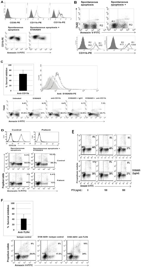
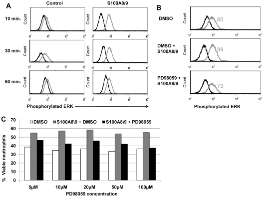
Programmed cell death (PCD) is a fundamental mechanism in tissue and cell homeostasis. It was long suggested that apoptosis regulates the cell number in diverse cell populations; however no clear mechanism was shown. Neutrophils are the short-lived, first-line defense of innate immunity, with an estimated t = 1/2 of 8 hours and a high turnover rate. Here we first show that spontaneous neutrophil constitutive PCD is regulated by cell concentrations. Using a proteomic approach, we identified the S100 A8/9 complex, which constitutes roughly 40% of cytosolic protein in neutrophils, as mediating this effect. We further demonstrate that it regulates cell survival via a signaling mechanism involving MEK-ERK via TLR4 and CD11B/CD18. This mechanism is suggested to have a fine-tuning role in regulating the neutrophil number in bone marrow, peripheral blood, and inflammatory sites.


Conflict of interest statement

Competing Interests: The authors have declared that no competing interests exist.

Figures

Figure 1
Figure 1. Constitutive spontaneous neutrophil PCD.
A. Kinetics of spontaneous constitutive neutrophil PCD. Neutrophils were isolated and allowed to undergo spontaneous constitutive PCD at a concentration of 1×10⁶/ml. Samples were obtained at the indicated intervals, and the apoptosis rate was measured using Annexin V-FITC and PI staining. B. Transmission electron microscopy (TEM) of spontaneous constitutive neutrophil PCD. Morphology of viable or early apoptotic (white arrow) and apoptotic (black arrows) neutrophils is shown. Apoptotic cells show the typical morphology of condensed cytoplasm and chromatin. Cells were prepared for TEM as described in Materials and methods. C. Inhibition of spontaneous constitutive neutrophil PCD by pan-caspase inhibitor zVAD-fmk. Sample dot plots of AnnexinV-PI staining of neutrophils undergoing spontaneous constitutive PCD for 14 h at a concentration of 1×10⁶/ml, in the presence or absence of 20 µM Zvad-fmk. Pan-caspase inhibition rescued cells from apoptotic death and viable cells are increased from 19 to 49% (p<0.001). Data are representative of 3 or more experiments.
Figure 2
Figure 2. Spontaneous constitutive neutrophil PCD is cell concentration-dependent.
A. Neutrophils underwent spontaneous constitutive PCD for 12 h at cell concentrations ranging from 0.5×10⁶/ml up to 50×106/ml. (The physiological blood concentration is 2−6×10⁶/ml.) Apoptosis was assessed by AnnexinV-FITC and PI staining, as described in Materials and methods. Data represents the mean ± SD of 3 experiments. B. Sample dot plots of AnnexinV-PI staining of neutrophils undergoing spontaneous constitutive PCD for 12 h in (a) 0.5×10⁶/ml, and (b) 16×106/ml. The percentage of viable, early (AnnexinV+/PI) and late (AnnexinV+/PI+) apoptotic cells is indicated within the respective quadrants. C. Survival of neutrophils undergoing spontaneous constitutive PCD, based on cell counts. Data presented is the mean ± SD of 3 experiments.
Figure 3
Figure 3. Identification of candidate molecules that induce the community effect.
A. Supernatant of cells undergoing spontaneous constitutive PCD at high concentrations has rescued cells undergoing spontaneous constitutive PCD from apoptosis at low concentrations, and improved their survival (p<0.001). Data is representative of 3 experiments. B. Representative dot plot of supernatant transfer assay. Apoptosis was assessed by AnnexinV and PI staining of neutrophils after 12 h of spontaneous constitutive PCD. The transfer assay was performed as follows: neutrophils underwent spontaneous constitutive PCD at low (a, 0.5×10⁶/ml) or high (c, 16×106/ml) concentration for 12 h. The supernatant of the cells incubated at high concentrations was collected and used as media for neutrophils undergoing constitutive spontaneous PCD at low concentrations (b), and the supernatant of the cells incubated at low concentrations was collected and used for neutrophils undergoing constitutive spontaneous PCD at high concentrations (d). C. SDS-PAGE of differentially displayed proteins from high- and low-concentration spontaneous constitutive PCD. The supernatants of cells undergoing spontaneous constitutive PCD at high- and low concentrations were collected, and the proteins were purified as described in Materials and methods. Protein samples were electrophorized and stained with Coomassie, as shown. Two proteins, ∼14 KD and ∼10 KD, were identified in the supernatant of cells undergoing spontaneous constitutive PCD at high concentrations (16×106/ml).
Figure 4
Figure 4. Effect of S100A9 and S100A8 on neutrophil apoptosis.
A. S100A9 and S100A8 add-in experiments. Varying concentrations of S100A9, S100A8, and the S100A8/9 complex were added to supernatants of neutrophils undergoing spontaneous constitutive PCD at a concentration of 0.5×10⁶/ml. After 10 hours apoptosis was evaluated using Annexin V-PI and mitochondrial staining. Data is presented as mean ± SD of Annexin V-PI staining (* p<0.05, ** p<0.02). B. S100A9 and S100A8 add-in experiments. A representative sample of Annexin V-PI and mitochondrial staining. Neutrophils undergoing spontaneous constitutive PCD without (a and c) or with (b and d) the addition of 2 µg/ml S100A8/9. Cells were stained either with Annexin V-FITC (a and b) or with DiOC6 (c and d), as well as PI (a, b, c, d) and assessed by flow cytometry. Dot plots are representative of 6 experiments. C. The effect of anti-S100A8 and A9 on add-in experiments. Neutrophils undergoing spontaneous constitutive PCD at a concentration of 0.5×10⁶/ml for 12h, with rabbit polyclonal antibody dilutions of 1∶100, 1∶1000, and 1∶10,000 against S100A9 and S100A8, were then treated with 2 µg/ml S100A8/9 complex. Rabbit serum at the same dilutions of 1∶100, 1∶1000, and 1∶10,000 was used as a control. Apoptosis was assessed using Annexin V-FITC and PI staining. Percentages of viable, early-, and late apoptotic cells are indicated within the respective quadrants. Dot plots are representative of 6 experiments.
Figure 5
Figure 5. The survival effect of S100A8/9 is mediated through CD11b/CD18 and TLR4.
A. Expression of CD11b on neutrophils. Upper panel. Freshly isolated neutrophils express CD11b/CD18 (isotype control is shown as filled histogram and anti- CD11b/CD18-PE in dotted line, middle) but not CD36 (left). Following 12 h of spontaneous PCD (gray line, middle) there was a marked decrease in the expression of CD11b/CD18 in comparison to freshly isolated neutrophils. Addition of S100A8/9 resulted in two different cell populations: high and low-CD11b/CD18 (black line, right). Lower panel. Left. Correlation between CD11b and phosphatidylserine expression demonstrates that when neutrophils become apoptotic they downregulate CD11b expression. Right. S100A8/9 upregulates CD11b expression. B. S100A8/9 dramatically increases CD11b on viable cells. Viable cells (R1, black line) increase CD11b expression from a mean fluorescence of 552 to 1094 (p<0.0001). Apoptotic cells (R2, gray line) still maintain some of this effect and show a mean fluorescence of 473, compared to 362 in the absence of S100A8/9. Filled histograms represent isotype control. Neutrophils were harvested after 12 h spontaneous constitutive PCD, with or without treatment with 2 µg/ml S100A8/9. Histograms are representative of at least 3 different experiments. C. The survival effect of S100A8/9 in the presence of anti-CD11b/CD18. Upper panel. Right. Neutrophils were incubated for 12 h and treated with either anti-CD11b or the isotype control IgG1 before addition of S100A8/9 complex. Integrin inhibition reduced the effect of S100A8/9 by 40−100% in comparison with the isotype control (p<0.001). The average of 6 experiments is presented. Lower panel. A representative dot plot is shown. Upper panel. Left. S100A8/9 binds to CD11b/CD18. CHO cells transfected with CR3 (black) or vector (gray) were evaluated for S100A8/9 binding. CR3-ransfected cells bound at rates almost twofold higher than CHO control cells (median fluorescence 13.3 vs. 6.6, p<0.001). The filled curve represents isotype control; the histogram is representative of 3 experiments. D. S100A8/9 effect on neutrophils from a CD11b/CD18-deficient patient. Neutrophil expression of CD11b from a healthy control and a patient with leukocyte adhesion deficiency is shown in the upper panel. Spontaneous constitutive apoptosis is shown in the lower panel, together with the S100A8/9 effect. Neutrophils were isolated from a CD11b-deficient patient and a healthy control, as described in Materials and methods, incubated at a concentration of 1×10⁶/ml and allowed to undergo spontaneous constitutive PCD for 10 h, with or without addition of S100A8/9. Apoptosis was assessed by Annexin V-PI staining. E. The survival effect induced by the complex S100A8/9 in the presence of pertussis toxin. Bordetella pertussis toxin at 100 and 500 ng/ml were added to neutrophils 45 min before adding S100A8/9. Neutrophils then were allowed to undergo constitutive spontaneous PCD that was assessed using AnnexinV-FITC and PI staining after 12 h. The dot plots are representative of 3 experiments. F. The survival effect of S100A8/9 in the presence of anti-TLR4. Neutrophils were incubated for 10 h and treated with either anti-TLR4 or the isotype control IgG2a, as described in Materials and methods. Inhibition of TLR4 abrogated the effect of S100A8/9 by 30−60% (p<0.001) in comparison with the isotype control, as seen in upper panel. The upper panel is presented as percentage of control. The experiment is a summary of four experiments (upper panel) with representative Plots in the lower panel.
Figure 6
Figure 6. Effects of the S100A8/9 complex are mediated through the MAPK-ERK pathway.
A. Phosphorylation of MAPK following exposure to S100A8/9. Freshly isolated neutrophils with or without treatment with 2 µg/ml S100A8/9 for various times as indicated, and then prepared for intracellular staining with mouse anti-human phospho-p44/42 MAPK alexa fluor 488 (gray) or isotype control (black), as described in Materials and methods. In the presence of S100A8/9, phosphorylation was increased by 3.22-fold after 10 min (p<0.001), 1.5-fold after 30 min (p<0.001), and decayed at 60 min (p<0.001). Histograms are representative of 3 independent experiments. B. Reduced phosphorylation of MAPK following exposure to S100A8/9 in the presence of PD98059, a MAPK phosphorylation inhibitor. Freshly isolated neutrophils were treated with the inhibitor PD98059 or with DMSO as a control for 30 min, and then treated with 2 µg/ml S100A8/9. Cells were then harvested and prepared for intracellular staining of phospho-p44/42 MAPK (gray) or isotype control (black), as described in Materials and methods. Treatment with PD98059 inhibitor reduced phosphorylation caused by the S100A8/9 complex (p<0.001). Histograms are representative of 3 experiments. C. Effect of the S100A8/9 complex in the presence of MAPK-ERK inhibitor. The effect of S100A8/9 was assessed in the presence of PD98059. Freshly isolated neutrophils were treated with varying combinations and concentrations of DMSO (white bars), S100A8/9 complex (gray bars), or S100A8/9 complex with PD98059 (black bars), as indicated, for 30 min before adding S100A8/9. Neutrophils were allowed to undergo constitutive spontaneous PCD. Results represent the percentage of viable cells according to Annexin V-FITC and PI staining. Data is representative of 3 experiments (p<0.001).

Similar articles

Cited by

References

    1. Ellis RE, Yuan JY, Horvitz HR. Mechanisms and functions of cell death. Annu Rev Cell Biol. 1991;7:663–698. - PubMed
    1. Krammer PH. CD95′s deadly mission in the immune system. Nature. 2000;407:789–795. - PubMed
    1. Dancey JT, Deubelbeiss KA, Harker LA, Finch CA. Neutrophil kinetics in man. J Clin Invest. 1976;58:705–715. - PMC - PubMed
    1. Metcalf D. Hematopoietic cytokines. Blood. 2008;111:485–491. - PMC - PubMed
    1. Stark MA, Huo Y, Burcin TL, Morris MA, Olson TS, et al. Phagocytosis of apoptotic neutrophils regulates granulopoiesis via IL-23 and IL-17. Immunity. 2005;22:285–294. - PubMed

Publication types

MeSH terms